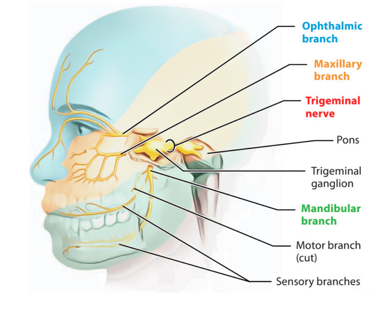
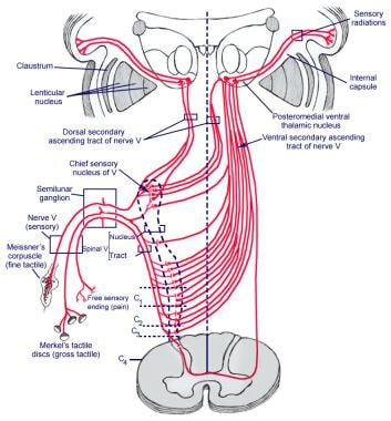
Trigeminal Nerve Pathway Diagram

Trigeminal Nerve вђ Anatomy Qa The peripheral aspect of the trigeminal ganglion gives rise to 3 divisions: ophthalmic (v1), maxillary (v2) and mandibular (v3). the motor root passes inferiorly to the sensory root, along the floor of the trigeminal cave. its fibres are only distributed to the mandibular division. the ophthalmic nerve and maxillary nerve travel lateral to the. The principal regulator of the sensory modalities of the head is the trigeminal nerve. this is the fifth of twelve pairs of cranial nerves that are responsible for transmitting numerous motor, sensory, and autonomous stimuli to structures of the head and neck. while the trigeminal nerve (cn v) is largely a sensory nerve, it also mingles in the.

Trigeminal Nerve вђ Earth S Lab Trigeminal nerve. the trigeminal nerve is the fifth cranial nerve. it is also represented as cn v. it is the largest of all the cranial nerves. it is the most complex of all the cranial nerves due to it's extensive anatomic course. this nerve is a mixed nerve having both sensory and motor fibres. Learn about the trigeminal nerve, the largest of the 12 cranial nerves that connects the face to the brain. see an interactive 3 d diagram of the nerve and its three branches, and find out how to test and protect it. The trigeminal nerve is the 5th cranial nerve (cn v) and the largest of the cranial nerves (see image. cranial nerves in the orbit). cn v provides most of the face's sensory innervation and the mastication muscles' motor stimulation.[1] the nerve's 3 main branches are the ophthalmic (v1), maxillary (v2), and mandibular (v3) nerves. these branches join at the trigeminal ganglia within the. The trigeminal nerve is responsible for carrying most of the sensation of the face to the brain. the sensory trigeminal nerve branches of the trigeminal nerve are the ophthalmic, the maxillary, and the mandibular nerves, which correspond to sensation in the v1, v2, and v3 regions of the face, respectively. ophthalmic nerve: this nerve detects.

Trigeminal Nerve Anatomy Gross Anatomy Branches Of The Trigeminal The trigeminal nerve is the 5th cranial nerve (cn v) and the largest of the cranial nerves (see image. cranial nerves in the orbit). cn v provides most of the face's sensory innervation and the mastication muscles' motor stimulation.[1] the nerve's 3 main branches are the ophthalmic (v1), maxillary (v2), and mandibular (v3) nerves. these branches join at the trigeminal ganglia within the. The trigeminal nerve is responsible for carrying most of the sensation of the face to the brain. the sensory trigeminal nerve branches of the trigeminal nerve are the ophthalmic, the maxillary, and the mandibular nerves, which correspond to sensation in the v1, v2, and v3 regions of the face, respectively. ophthalmic nerve: this nerve detects. Figure 1: divisions of the trigeminal nerve (cn v), lateral view. figure 2: branches of the ophthalmic nerve (cn v1), parasagittal view. figure 3: branches of the maxillary nerve (cn v2), a. parasagittal view, and b. midsagittal view with bony nasal septum removed. figure 4: branches of the mandibular nerve (cn v3), parasagittal view. The motor root of the trigeminal nerve bypasses the trigeminal ganglion and reunites with the mandibular nerve in the foramen ovale basis cranii . as the mandibular nerve enters the masticator space, it divides into several sensory branches to supply sensation to the lower third of the face and the tongue, floor of the mouth, and the jaw ( fig.

Schematic Drawing Of The Trigeminal Nerve Download Scientific Diagram Figure 1: divisions of the trigeminal nerve (cn v), lateral view. figure 2: branches of the ophthalmic nerve (cn v1), parasagittal view. figure 3: branches of the maxillary nerve (cn v2), a. parasagittal view, and b. midsagittal view with bony nasal septum removed. figure 4: branches of the mandibular nerve (cn v3), parasagittal view. The motor root of the trigeminal nerve bypasses the trigeminal ganglion and reunites with the mandibular nerve in the foramen ovale basis cranii . as the mandibular nerve enters the masticator space, it divides into several sensory branches to supply sensation to the lower third of the face and the tongue, floor of the mouth, and the jaw ( fig.

Comments are closed.