Trigeminal Nerve вђ Anatomy Qa

Trigeminal Nerve вђ Anatomy Qa It is a pure sensory nerve. it arises from the anterior aspect of trigeminal ganglion. it then passes forward along the lateral wall of cavernous sinus. it divides into three branches – nasocilliary, frontal and lacrimal. the three branches exit cranial cavity by passing through the superior orbital fissure. The principal regulator of the sensory modalities of the head is the trigeminal nerve. this is the fifth of twelve pairs of cranial nerves that are responsible for transmitting numerous motor, sensory, and autonomous stimuli to structures of the head and neck. while the trigeminal nerve (cn v) is largely a sensory nerve, it also mingles in the.

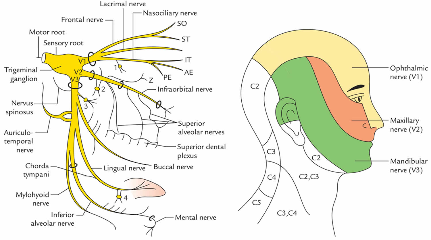
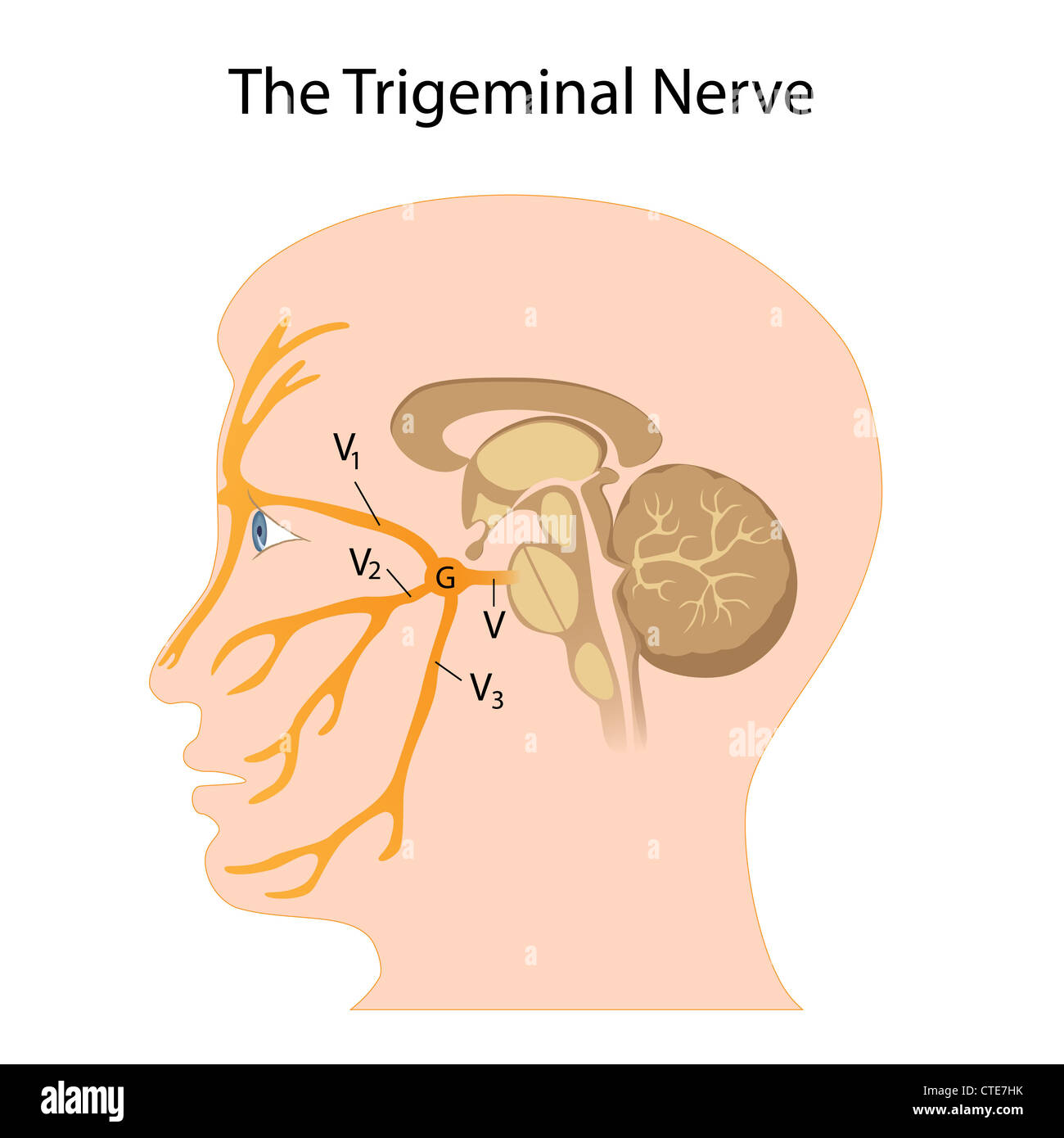
Trigeminal Nerve Anatomy Branches Distribution Function Damage Pain The peripheral aspect of the trigeminal ganglion gives rise to 3 divisions: ophthalmic (v1), maxillary (v2) and mandibular (v3). the motor root passes inferiorly to the sensory root, along the floor of the trigeminal cave. its fibres are only distributed to the mandibular division. the ophthalmic nerve and maxillary nerve travel lateral to the. Content:0:00 introduction01:05 trigeminal nerve scheme08:19 trigeminal nerve nucleus09:57 course of the trigeminal nerve10:49 ophthalmic nerve (v1)14:07 maxi. When it comes to the trigeminal nerve, or cn v, tri means three, so right off the bat, you can tell that the trigeminal nerve has three major branches: the ophthalmic nerve, or v1, the maxillary nerve, or v2, and the mandibular nerve, or v3. the ophthalmic and the maxillary nerves only have sensory functions, while the mandibular nerve has both. The trigeminal nerve is the 5th cranial nerve (cn v) and the largest of the cranial nerves (see image. cranial nerves in the orbit). cn v provides most of the face's sensory innervation and the mastication muscles' motor stimulation.[1] the nerve's 3 main branches are the ophthalmic (v1), maxillary (v2), and mandibular (v3) nerves. these branches join at the trigeminal ganglia within the.

Trigeminal Nerve Anatomy When it comes to the trigeminal nerve, or cn v, tri means three, so right off the bat, you can tell that the trigeminal nerve has three major branches: the ophthalmic nerve, or v1, the maxillary nerve, or v2, and the mandibular nerve, or v3. the ophthalmic and the maxillary nerves only have sensory functions, while the mandibular nerve has both. The trigeminal nerve is the 5th cranial nerve (cn v) and the largest of the cranial nerves (see image. cranial nerves in the orbit). cn v provides most of the face's sensory innervation and the mastication muscles' motor stimulation.[1] the nerve's 3 main branches are the ophthalmic (v1), maxillary (v2), and mandibular (v3) nerves. these branches join at the trigeminal ganglia within the. Figure 2.brainstem nuclei. 2 the trigeminal nerve roots. both motor and sensory components of the trigeminal nerve complex exit the ventral mid pons as distinct nerves the larger, more medial nerve is the trigeminal sensory root; and a smaller, more lateral nerve is the trigeminal motor root named portio minor (the minor portion of the trigeminal nerve; the fourth branch). E. in neuroanatomy, the trigeminal nerve (lit. triplet nerve), also known as the fifth cranial nerve, cranial nerve v, or simply cn v, is a cranial nerve responsible for sensation in the face and motor functions such as biting and chewing; it is the most complex of the cranial nerves. its name (trigeminal, from latin tri 'three' and geminus.

Trigeminal Nerve Anatomy Mri Figure 2.brainstem nuclei. 2 the trigeminal nerve roots. both motor and sensory components of the trigeminal nerve complex exit the ventral mid pons as distinct nerves the larger, more medial nerve is the trigeminal sensory root; and a smaller, more lateral nerve is the trigeminal motor root named portio minor (the minor portion of the trigeminal nerve; the fourth branch). E. in neuroanatomy, the trigeminal nerve (lit. triplet nerve), also known as the fifth cranial nerve, cranial nerve v, or simply cn v, is a cranial nerve responsible for sensation in the face and motor functions such as biting and chewing; it is the most complex of the cranial nerves. its name (trigeminal, from latin tri 'three' and geminus.

Comments are closed.