The Mouth And Buccal Cavity Anatomy Of The Human Mouth

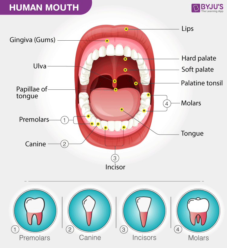
The Mouth And Buccal Cavity Anatomy Of The Human Mouth The oral cavity, or more commonly known as the mouth or buccal cavity, serves as the first portion of the digestive system. it consists of several different anatomically different aspects that work together effectively and efficiently to perform several functions. these aspects include the lips, tongue, palate, and teeth. although a small compartment, the oral cavity is a unique and complex. The buccal cavity mainly comprises the primary organ of the digestive system including the teeth, tongue and salivary glands. also, read digestive system in humans. the mouth is an opening through which the food is taken inside the body. it is bounded by lips and its inner parts comprise the cheeks, tongue, upper jaw and lower jaw.

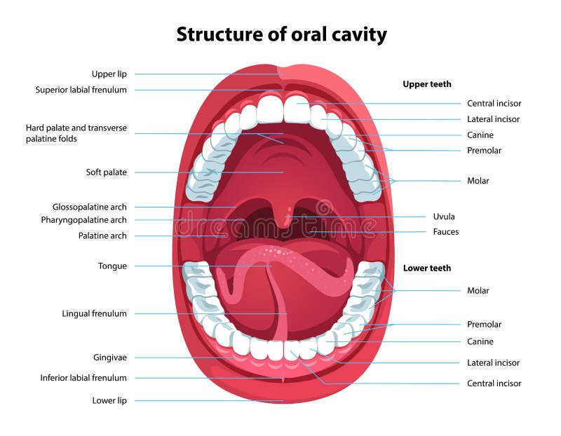
Structure Of Oral Cavity Human Mouth Anatomy Stock Vector Lips. mouth, in human anatomy, orifice through which food and air enter the body. the mouth opens to the outside at the lips and empties into the throat at the rear; its boundaries are defined by the lips, cheeks, hard and soft palates, and glottis. it is divided into two sections: the vestibule, the area between the cheeks and the teeth, and. The oral cavity, better known as the mouth, is the start of the alimentary canal. it has three major functions: digestion – receives food, preparing it for digestion in the stomach and small intestine. communication – modifies the sound produced in the larynx to create a range of sounds. breathing – acts as an air inlet in addition to the. Oral cavity. the oral cavity is situated anteriorly on the face, under the nasal cavities.it is bounded by a roof, a floor and lateral walls. anteriorly it opens to the face through the oral fissure, while posteriorly the oral cavity communicates with the oropharynx through a narrow passage called the oropharyngeal isthmus (also termed the isthmus of the fauces). Human mouth. in human anatomy, the mouth is the first portion of the alimentary canal that receives food and produces saliva. [2] the oral mucosa is the mucous membrane epithelium lining the inside of the mouth. in addition to its primary role as the beginning of the digestive system, the mouth also plays a significant role in communication.

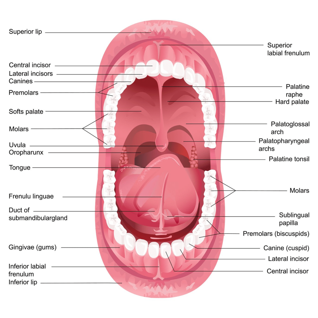
Mouth Buccal Cavity And Tongue Neet Notes Edurev Oral cavity. the oral cavity is situated anteriorly on the face, under the nasal cavities.it is bounded by a roof, a floor and lateral walls. anteriorly it opens to the face through the oral fissure, while posteriorly the oral cavity communicates with the oropharynx through a narrow passage called the oropharyngeal isthmus (also termed the isthmus of the fauces). Human mouth. in human anatomy, the mouth is the first portion of the alimentary canal that receives food and produces saliva. [2] the oral mucosa is the mucous membrane epithelium lining the inside of the mouth. in addition to its primary role as the beginning of the digestive system, the mouth also plays a significant role in communication. The cheeks, tongue, and palate frame the mouth, which is also called the oral cavity (or buccal cavity). the structures of the mouth are illustrated in figure 23.3.1. at the entrance to the mouth are the lips, or labia (singular = labium). their outer covering is skin, which transitions to a mucous membrane in the mouth proper. We have created 114 medical original illustrations of the mouth, the buccal cavity, the bones of the palate, the tongue, the salivary glands and the oral part of the pharynx with vessels and nerves. the drawing and anatomical labeling (using the terminologia anatomica 2) of these illustrations was done by gauthier kervyn under the anatomical.

Mouth And Teeth Diagram The cheeks, tongue, and palate frame the mouth, which is also called the oral cavity (or buccal cavity). the structures of the mouth are illustrated in figure 23.3.1. at the entrance to the mouth are the lips, or labia (singular = labium). their outer covering is skin, which transitions to a mucous membrane in the mouth proper. We have created 114 medical original illustrations of the mouth, the buccal cavity, the bones of the palate, the tongue, the salivary glands and the oral part of the pharynx with vessels and nerves. the drawing and anatomical labeling (using the terminologia anatomica 2) of these illustrations was done by gauthier kervyn under the anatomical.

Comments are closed.