Spinal Nerves Labelled On Lacc Model Nerve Anatomy Anatomy Modelsођ

Spinal Nerves Model From Lacc Spinal Cord Anatomy Nerve St Sciatic n. what is a? common fibular division. what is b? tibial division. what is c? study with quizlet and memorize flashcards containing terms like posterior ramus, posterior root, anterior root and more. Myelin sheath. schwann cell. node of ranvier. endoneurium. (wraps the schwann cells) perineurium. (wraps the fascicle) fascicle. study with quizlet and memorize flashcards containing terms like cervical plexus, brachial plexus, lumbosacral plexus and more.

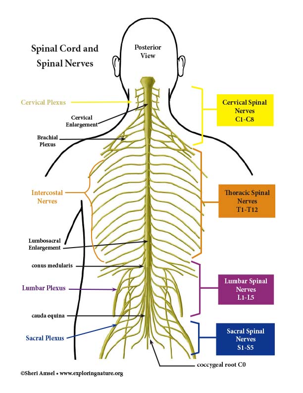
Spinal Nerve Anatomy Diagram Anatomy of spinal cord. extends from base of brain to between l1 or l2. crosses vertebral foramen. spinal nerves cross intervertebral foramina. spinal cord shorter than vertebral column in adults stops growing at age 4. spinal nerves enter vertebral column at a point that is caudal to their insertion points in the spinal cord. Function. receive sensory information from the periphery and pass them to the cns. recieve motor information from the cns and pass them to the periphery. clinical relations. nerve root impingement, disk protrusion, disk herniation, spinal stenosis, spinal nerve impingement. this article will discuss the anatomy and function of the spinal nerves. Spinal nerve. medically reviewed by anatomy team. spinal nerves are mixed nerves that emerge from the spinal cord and carry both motor and sensory information between the spinal cord and various parts of the body. these nerves are essential for transmitting sensory signals to the brain and for carrying motor commands from the brain to muscles. These allow us to control the many muscles in our bodies. the spinal nerves are divided into four main categories of spinal nerves based on the location from which they branch. 8 cervical (c1 c8) nerves emerge from the cervical spine (neck) 12 thoracic (t1 t12) nerves emerge from the thoracic spine (mid back) 5 lumbar (l1 l5) nerves emerge from.

Anatomy Chart Spinal Nerves Spinal nerve. medically reviewed by anatomy team. spinal nerves are mixed nerves that emerge from the spinal cord and carry both motor and sensory information between the spinal cord and various parts of the body. these nerves are essential for transmitting sensory signals to the brain and for carrying motor commands from the brain to muscles. These allow us to control the many muscles in our bodies. the spinal nerves are divided into four main categories of spinal nerves based on the location from which they branch. 8 cervical (c1 c8) nerves emerge from the cervical spine (neck) 12 thoracic (t1 t12) nerves emerge from the thoracic spine (mid back) 5 lumbar (l1 l5) nerves emerge from. Figure 1. rami of a spinal nerve. in the thoracic region, the ventral rami of spinal nerves t2 t12 form intercostal nerves. figure 2. nerve plexuses of the body there are four main nerve plexuses in the human body. the cervical plexus supplies nerves to the posterior head and neck, as well as to the diaphragm. The cervical plexus is composed of axons from spinal nerves c 1 through c 5. it branches into nerves in the posterior neck and head as well as the phrenic nerve, which connects to the diaphragm at the base of the thoracic cavity. figure 12.4.5 12.4. 5: nerve plexuses of the body.

Spinal Nerves Labelled On Lacc Model Nerve Anatomy Anat Figure 1. rami of a spinal nerve. in the thoracic region, the ventral rami of spinal nerves t2 t12 form intercostal nerves. figure 2. nerve plexuses of the body there are four main nerve plexuses in the human body. the cervical plexus supplies nerves to the posterior head and neck, as well as to the diaphragm. The cervical plexus is composed of axons from spinal nerves c 1 through c 5. it branches into nerves in the posterior neck and head as well as the phrenic nerve, which connects to the diaphragm at the base of the thoracic cavity. figure 12.4.5 12.4. 5: nerve plexuses of the body.

Comments are closed.