Spinal Cord Anatomy Wikimsk Vrogue Co

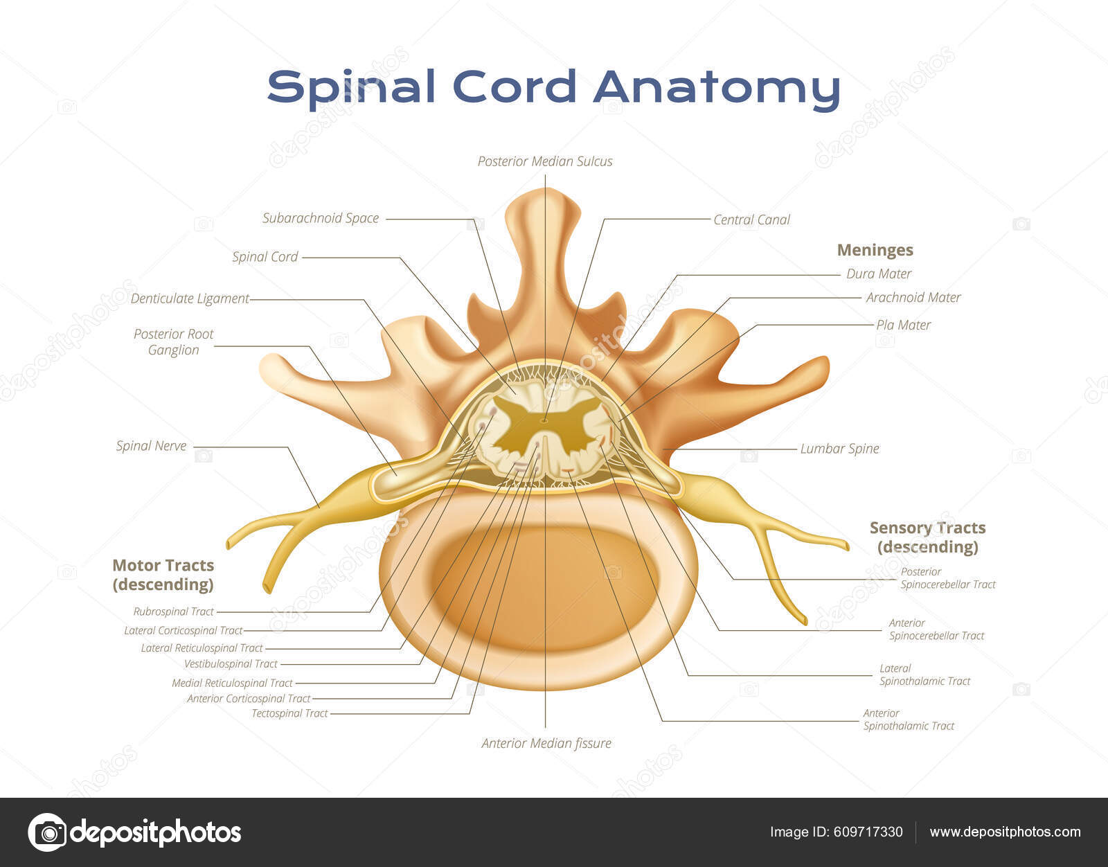
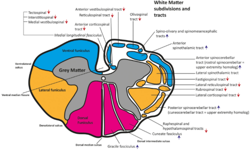
Spinal Cord Anatomy Wikimsk Spinal cord sectional anatomy. a deep ventral median fissure and a shallower dorsal median sulcus and septum posteriorly divides the spinal cord into right and left halves that are almost completely separated. dorsal nerve roots travel to the cord on the posterior surface of the cord through the dorsolateral sulci. The spinal cord is part of the central nervous system (cns). it is situated inside the vertebral canal of the vertebral column. during development, there’s a disproportion between spinal cord growth and vertebral column growth. the spinal cord finishes growing at the age of 4, while the vertebral column finishes growing at age 14 18.

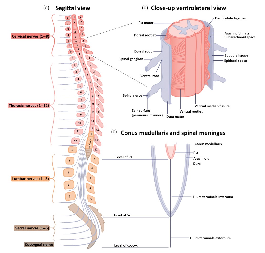
The Spinal Cord And Its Importance Spinal Cord Anatom Vrogue Co Sectional organization of spinal cord. the spinal cord is the main pathway for information connecting the brain and peripheral nervous system. [3] [4] much shorter than its protecting spinal column, the human spinal cord originates in the brainstem, passes through the foramen magnum, and continues through to the conus medullaris near the second lumbar vertebra before terminating in a fibrous. The spinal cord study is one of the most complex yet quite a fascinating part of the nervous system. its complex connections, the development defects, the lesions, and clinical presentation are quite overwhelming and warrants a better understanding of its anatomical and physiological nature. this topic has received extensive study and revealed many minute details. but it quite acknowledgeable. The spinal cord is a tubular bundle of nervous tissue and supporting cells that extends from the brainstem to the lumbar vertebrae. together, the spinal cord and the brain form the central nervous system. in this article, we shall examine the macroscopic anatomy of the spinal cord – its structure, membranous coverings and blood supply. The spinal cord is a long, tube like band of tissue. it connects your brain to your lower back. your spinal cord carries nerve signals from your brain to your body and vice versa. these nerve signals help you feel sensations and move your body. any damage to your spinal cord can affect your movement or function.

Vertebrae Spinal Cord Anatomy Infographics With Scien Vrogue Co The spinal cord is a tubular bundle of nervous tissue and supporting cells that extends from the brainstem to the lumbar vertebrae. together, the spinal cord and the brain form the central nervous system. in this article, we shall examine the macroscopic anatomy of the spinal cord – its structure, membranous coverings and blood supply. The spinal cord is a long, tube like band of tissue. it connects your brain to your lower back. your spinal cord carries nerve signals from your brain to your body and vice versa. these nerve signals help you feel sensations and move your body. any damage to your spinal cord can affect your movement or function. The spinal cord sits inside the spinal or vertebral canal. this canal also contains the meninges (membranes that protect the spinal cord), blood vessels, spinal nerve roots, and surrounding fatty and connective tissues. the spinal canal is formed by the vertebral foramina of the vertebral bodies. the anterior border of the canal is made up of. Anatomy. the spinal cord is a mass of nervous tissue that extends inferiorly from the brain stem through the vertebral canal of the cervical and thoracic regions, ending around the t12 or l1 vertebra. it is a long tube about 18 inches (45 cm) in length and around half an inch (1 cm) in diameter at its widest point.

Spinal Cord Anatomy Wikimsk The spinal cord sits inside the spinal or vertebral canal. this canal also contains the meninges (membranes that protect the spinal cord), blood vessels, spinal nerve roots, and surrounding fatty and connective tissues. the spinal canal is formed by the vertebral foramina of the vertebral bodies. the anterior border of the canal is made up of. Anatomy. the spinal cord is a mass of nervous tissue that extends inferiorly from the brain stem through the vertebral canal of the cervical and thoracic regions, ending around the t12 or l1 vertebra. it is a long tube about 18 inches (45 cm) in length and around half an inch (1 cm) in diameter at its widest point.

Comments are closed.