Spinal Cord Anatomy Wikimsk

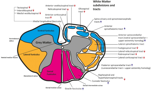
Spinal Cord Anatomy Wikimsk Spinal cord sectional anatomy. a deep ventral median fissure and a shallower dorsal median sulcus and septum posteriorly divides the spinal cord into right and left halves that are almost completely separated. dorsal nerve roots travel to the cord on the posterior surface of the cord through the dorsolateral sulci. Main article: spinal cord anatomy. part or all of this article or section is derived from numbness by ddxof, used under cc by sa. sensory information is gathered from specialized receptors in skin and soft tissues which detect a variety of stimuli including temperature, pressure, vibration, and pain. this is subsequently transmitted through.

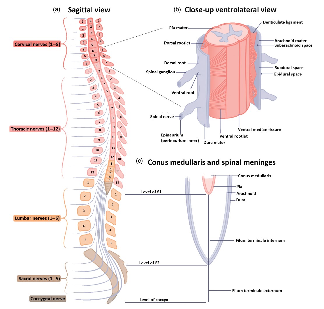
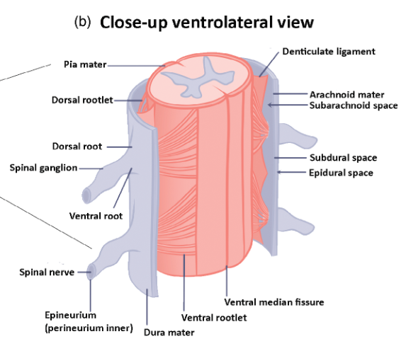
Spinal Cord Anatomy Wikimsk Dermatomes vary based on the method of assessment, the type of sensation tested, and the precise neural element being tested. lee et al's map is the most consistent for tactile dermatomal regions for each spinal dorsal nerve root. the midline has minimal overlap, but otherwise there is extensive and variable overlap. Spinal cord. the spinal cord is a long, thin, tubular structure made up of nervous tissue that extends from the medulla oblongata in the brainstem to the lumbar region of the vertebral column (backbone) of vertebrate animals. the center of the spinal cord is hollow and contains a structure called the central canal, which contains cerebrospinal. The spinal cord study is one of the most complex yet quite a fascinating part of the nervous system. its complex connections, the development defects, the lesions, and clinical presentation are quite overwhelming and warrants a better understanding of its anatomical and physiological nature. this topic has received extensive study and revealed many minute details. but it quite acknowledgeable. The spinal cord is a tubular bundle of nervous tissue and supporting cells that extends from the brainstem to the lumbar vertebrae. together, the spinal cord and the brain form the central nervous system. in this article, we shall examine the macroscopic anatomy of the spinal cord – its structure, membranous coverings and blood supply.

Spinal Cord Anatomy Wikimsk The spinal cord study is one of the most complex yet quite a fascinating part of the nervous system. its complex connections, the development defects, the lesions, and clinical presentation are quite overwhelming and warrants a better understanding of its anatomical and physiological nature. this topic has received extensive study and revealed many minute details. but it quite acknowledgeable. The spinal cord is a tubular bundle of nervous tissue and supporting cells that extends from the brainstem to the lumbar vertebrae. together, the spinal cord and the brain form the central nervous system. in this article, we shall examine the macroscopic anatomy of the spinal cord – its structure, membranous coverings and blood supply. Csf is an ultrafiltrate of blood plasma through the permeable capillaries of the choroid plexus. volume. total csf volume between brain, spinal cord, and thecal sac is ~150 ml. csf formation occurs at rate of ~500 ml per day. thus, the total amount of csf is turned over 3 4 times per day. nerve root anatomy. cervical spine. Anatomy. the spinal cord is a mass of nervous tissue that extends inferiorly from the brain stem through the vertebral canal of the cervical and thoracic regions, ending around the t12 or l1 vertebra. it is a long tube about 18 inches (45 cm) in length and around half an inch (1 cm) in diameter at its widest point.

Comments are closed.