Scanning Electron Microscopy Sem Operation Image Analysis Video Jove

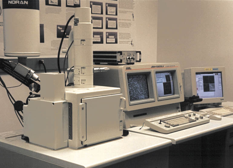
Scanning Electron Microscopy Sem Operation Image Analysis Video Jove A scanning electron microscope, or sem, is a powerful microscope that uses electrons to form an image. it allows for imaging of conductive samples at magnifications that cannot be achieved using traditional microscopes. modern light microscopes can achieve a magnification of ~1,000x, while typical sem can reach magnifications of more than 30,000x. Scanning electron microscopy, or sem, is a powerful technique used in chemistry and material analysis that uses a scanned electron beam to analyze the surface structure and chemical composition of a sample. modern light microscopes are limited by the interaction of visible light waves with an object, called diffraction.

Scanning Electron Microscopy Sem A scanning electron microscope, or sem, is a powerful microscope that uses electrons to form an image. it allows for imaging of conductive samples at magnifications that cannot be achieved using traditional microscopes. modern light microscopes can achieve a magnification of ~1,000x, while typical sem can reach magnifications of more than 30,000x. The sample was then inserted into the focused ion beam scanning electron microscope for three dimensional imaging. the focused ion beam was then used to sequentially remove thin layers of the sample. each layer was imaged prior to removal using backscatter sem. you’ve just watched jove’s introduction to scanning electron microscopy. Source: laboratory of dr. andrew j. steckl — university of cincinnati a scanning electron microscope, or sem, is a powerful microscope that uses. The scanning electron microscope works on the principle of applying kinetic energy to produce signals on the interaction of the electrons. these electrons are secondary electrons, backscattered electrons, and diffracted backscattered electrons which are used to view crystallized elements and photons. secondary and backscattered electrons are.

Scanning Electron Microscopy Sem Operation Image Analysis Source: laboratory of dr. andrew j. steckl — university of cincinnati a scanning electron microscope, or sem, is a powerful microscope that uses. The scanning electron microscope works on the principle of applying kinetic energy to produce signals on the interaction of the electrons. these electrons are secondary electrons, backscattered electrons, and diffracted backscattered electrons which are used to view crystallized elements and photons. secondary and backscattered electrons are. Purpose –the fei quanta feg 650 scanning electron microscope is a variable pressure microscope capable of resolving features at a scale of ~5 nm on samples up to 6 inch in size. the sem is equipped with 8 detectors for imaging and analysis. this document describes important information and basic operation procedure for imaging. Read this jove article on visualizing membrane ruffle formation using scanning electron microscopy. jove. jove. 교수 리소스 센터.

Comments are closed.