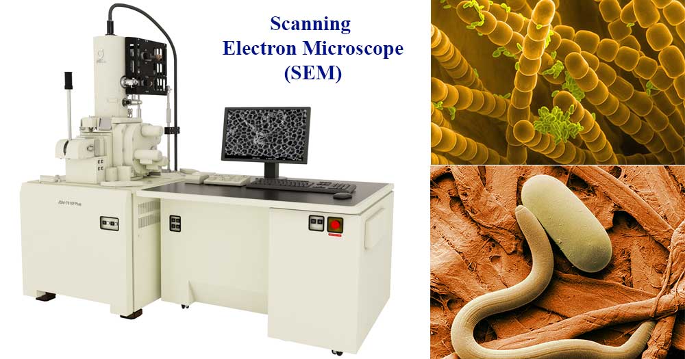
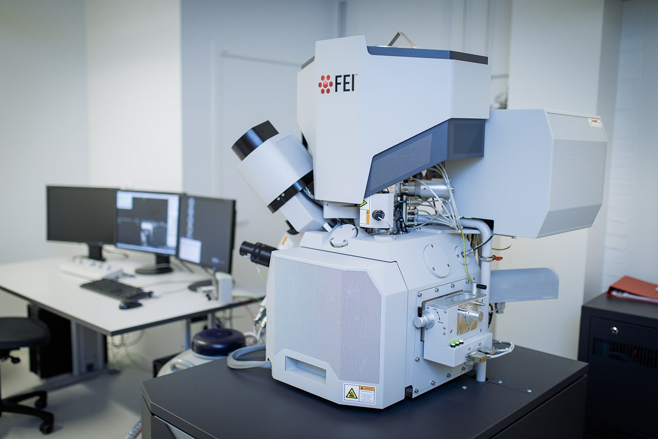
Scanning Electron Microscope Sem

Scanning Electron Microscope Sem Principle Parts Uses Microbe Notes A scanning electron microscope (sem) is a type of electron microscope that produces images of a sample by scanning the surface with a focused beam of electrons. the electrons interact with atoms in the sample, producing various signals that contain information about the surface topography and composition of the sample. Scanning electron microscope (sem) is a type of electron microscope that scans surfaces of microorganisms that uses a beam of electrons moving at low energy to focus and scan specimens. the development of electron microscopes was due to the inefficiency of the wavelength of light microscopes. electron microscopes have very short wavelengths in.

Scanning Electron Microscope Sem Definition Images Uses Scanning electron microscope (sem), type of electron microscope, designed for directly studying the surfaces of solid objects, that utilizes a beam of focused electrons of relatively low energy as an electron probe that is scanned in a regular manner over the specimen. the electron source and electromagnetic lenses that generate and focus the. Manfred von ardenne developed the first version of the sem in 1937. q5. what is the cost of a scanning electron microscope? the price of a new electron microscope ranges between $80,000 to $10,000,000 and above depending on the customizations, configurations, resolution, components, and brand value. Scanning electron microscopy (sem) in sem, the electron beam scans the sample in a raster pattern. instead of passing through the specimen, electrons get reflected on the surface or even ionize atoms within the sample by liberating electrons. these so called secondary electrons, as well as the backscattered electrons,. That’s the basic principle, but let’s dive a bit deeper into how the sem actually works. scanning electron microscopes have an electron gun, condenser and objective lenses, condenser and objective apertures, scan coils, and electron detectors. all of this also needs to be in a vacuum chamber so the electrons are not disturbed by gas molecules.

Scanning Electron Microscope Sem Working Principle Parts Scanning electron microscopy (sem) in sem, the electron beam scans the sample in a raster pattern. instead of passing through the specimen, electrons get reflected on the surface or even ionize atoms within the sample by liberating electrons. these so called secondary electrons, as well as the backscattered electrons,. That’s the basic principle, but let’s dive a bit deeper into how the sem actually works. scanning electron microscopes have an electron gun, condenser and objective lenses, condenser and objective apertures, scan coils, and electron detectors. all of this also needs to be in a vacuum chamber so the electrons are not disturbed by gas molecules. The electron beam of a scanning electron microscope interacts with atoms at different depths within the sample to produce different signals including secondary electrons, back scattered electrons, and characteristic x rays. each of these signals has its own detector in the sem, as seen in figure 1. secondary electrons are low energy electrons. Scanning electron microscopy is a characterization technique that images and analyzes a specimen by scanning an accelerated electron beam, followed by selectively collecting and recording secondary electrons, back scattered electrons, and other signals arising from the beam and specimen interactions. a modern scanning electron microscope (sem.

Comments are closed.