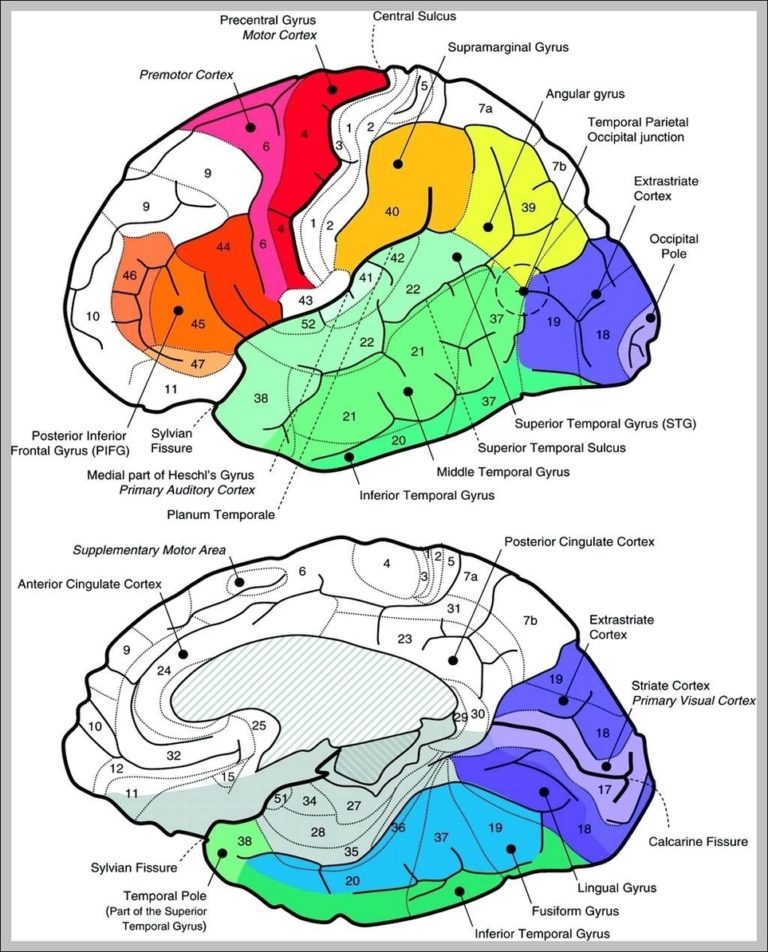
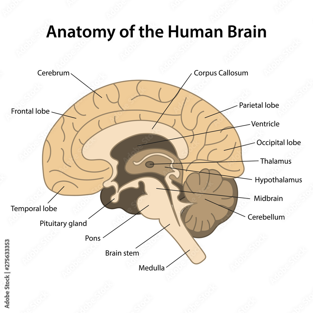
Sagittal View Of The Human Brain Image Anatomy System Human Body

Sagittal View Of The Human Brain Image Anatomy System Human Body Anatomical planes are imaginary planes 2d surfaces used to divide the body to facilitate descriptions of location and movement. the anatomical position is used as a reference when describing locations of structures and movements. it is an upright position with arms by the side and palms facing forward. feet are parallel with toes facing forward. This body anatomy diagram is great for learning about human health, is best for medical students, kids and general education. sagittal view of the human brain image the sagital view of the brain reflects some of the inverted c shaped rings including the cortex, cingulate gyrus, indusium griseum, corpus callosum, septum pellucidum lateral ventricles, and the thalamus which is central.

Anatomy Of The Human Brain Sagittal Cut Structure Of The Human Brai Explore the structure and function of the human brain in 3d, with interactive models, videos, and articles on brainfacts.org. The human brain is often sectioned (cut) and viewed from different directions and angles. 1. 2. previous. each point of view provides an altered perspective of the brain that changes the appearance of the major divisions, landmarks, and structures. The reticular formation is located throughout the brainstem. networks within the reticular formation are important for regulating sleep and consciousness, pain, and motor control. the fourth ventricle lies between the brainstem and the cerebellum. figure 18.2. regions of the diencephalon and brainstem in a midsagittal section. These anatomical charts include the main diagrams necessary for medical students, nursing students, residents, practitioners, anatomists to study the anatomy of the brain, to illustrate a course or explain a pathology to a patient. the basic structure of a neuron and an overall diagram of the human nervous system.

Sagital Section Of The Human Brain With Regions And Labels Stock Photo The reticular formation is located throughout the brainstem. networks within the reticular formation are important for regulating sleep and consciousness, pain, and motor control. the fourth ventricle lies between the brainstem and the cerebellum. figure 18.2. regions of the diencephalon and brainstem in a midsagittal section. These anatomical charts include the main diagrams necessary for medical students, nursing students, residents, practitioners, anatomists to study the anatomy of the brain, to illustrate a course or explain a pathology to a patient. the basic structure of a neuron and an overall diagram of the human nervous system. Nomenclature the nomenclature is a collection of all terms used in all atlases and provides the consistent abbreviations used in the atlas of the human brain. once you have specified a structure you can use the nomenclature in the database section to look up the same region in other atlases. for german students we developed a glossary on our. The human brain atlas. in this atlas you can view mri sections through a living human brain as well as corresponding sections stained for cell bodies or for nerve fibers. the stained sections are from a different brain than the one which was scanned for the mri images. furthermore, for the stained sections, the brain was removed from the skull.

Comments are closed.