Rheumatoid Arthritis Hands X Ray

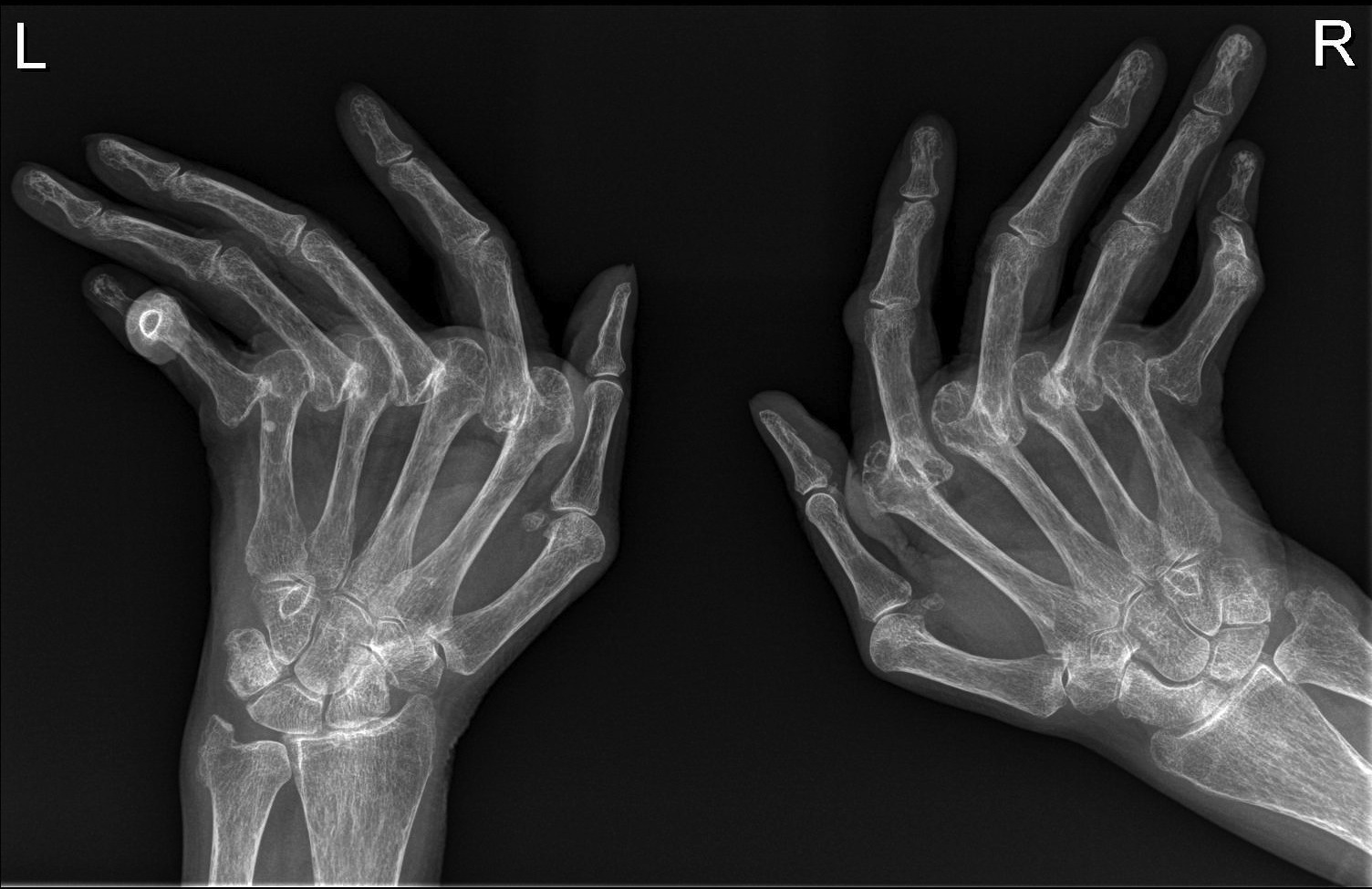
Rheumatoid Arthritis Hands Radiology At St Vincent S University Rheumatoid arthritis is a synovial based process, with a predilection for the: proximal interphalangeal and metacarpophalangeal joints (especially those of the index and middle fingers) ulnar styloid. triquetrum. as a rule, the distal interphalangeal joints are spared. late changes include:. The 2010 acr eular classification criteria for rheumatoid arthritis 4 has a maximal score of 10 and requires a score of >6 for a diagnosis of rheumatoid arthritis to be made: joint involvement. 0: 1 large joint. 1: 2 10 large joints. 2: 1 3 small joints (with or without the involvement of large joints).

Rheumatoid Arthritis X Ray Wikidoc Symmetrical pattern. rheumatoid arthritis manifests as a symmetrical arthritis, most commonly affecting the hands. if the pattern of disease is not symmetrical, then a different diagnosis should be considered. in early rheumatoid there may be no changes visible on an x ray. ultrasound can be used to look for erosions, synovitis, and tenosynovitis. Rheumatoid arthritis (ra) imaging tests are used to look for signs of ra and to monitor the disease’s progression. these tests primarily look for bone damage in the patient’s joints caused by the inflammation associated with ra. x rays used to be the most common form of imaging ordered, but they…. Updated april 28, 2022. for decades, x rays were used to help detect rheumatoid arthritis (ra) and monitor for worsening bone damage. in the early stages of ra, however, x rays may appear normal although the disease is active, making the films useful as a baseline but not much help in getting a timely diagnosis and treatment. Rheumatoid arthritis (ra) is an immune mediated multisystem inflammatory disease that predominantly affects the synovial joints. it was first described by alfred baring garrod in the year 1800. the disease can lead to inflammation, joint destruction, deformity, and disability, and may also present with extra articular manifestations. inflammatory arthritis involving the small joints of the.

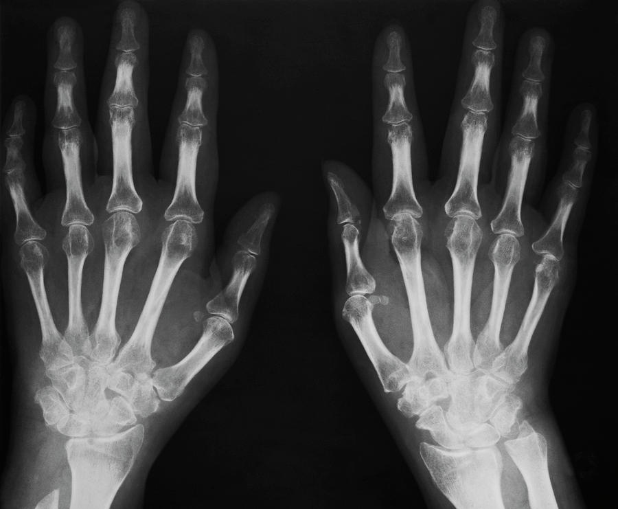
Arthritic Hands X Ray Photograph By Antonia Reeve Science Photo Library Updated april 28, 2022. for decades, x rays were used to help detect rheumatoid arthritis (ra) and monitor for worsening bone damage. in the early stages of ra, however, x rays may appear normal although the disease is active, making the films useful as a baseline but not much help in getting a timely diagnosis and treatment. Rheumatoid arthritis (ra) is an immune mediated multisystem inflammatory disease that predominantly affects the synovial joints. it was first described by alfred baring garrod in the year 1800. the disease can lead to inflammation, joint destruction, deformity, and disability, and may also present with extra articular manifestations. inflammatory arthritis involving the small joints of the. Introduction. rheumatoid arthritis (ra) is a symmetric, inflammatory, peripheral polyarthritis of unknown etiology. it typically leads to joint destruction through the erosion of cartilage and bone. untreated, it will lead to loss of physical function, inability to carry out daily tasks of living, and difficulties in maintaining employment. X ray images can help diagnose rheumatoid arthritis by showing changes in your bones and joints. ezzati f, et al. (2022). radiographic findings of inflammatory arthritis and mimics in the.

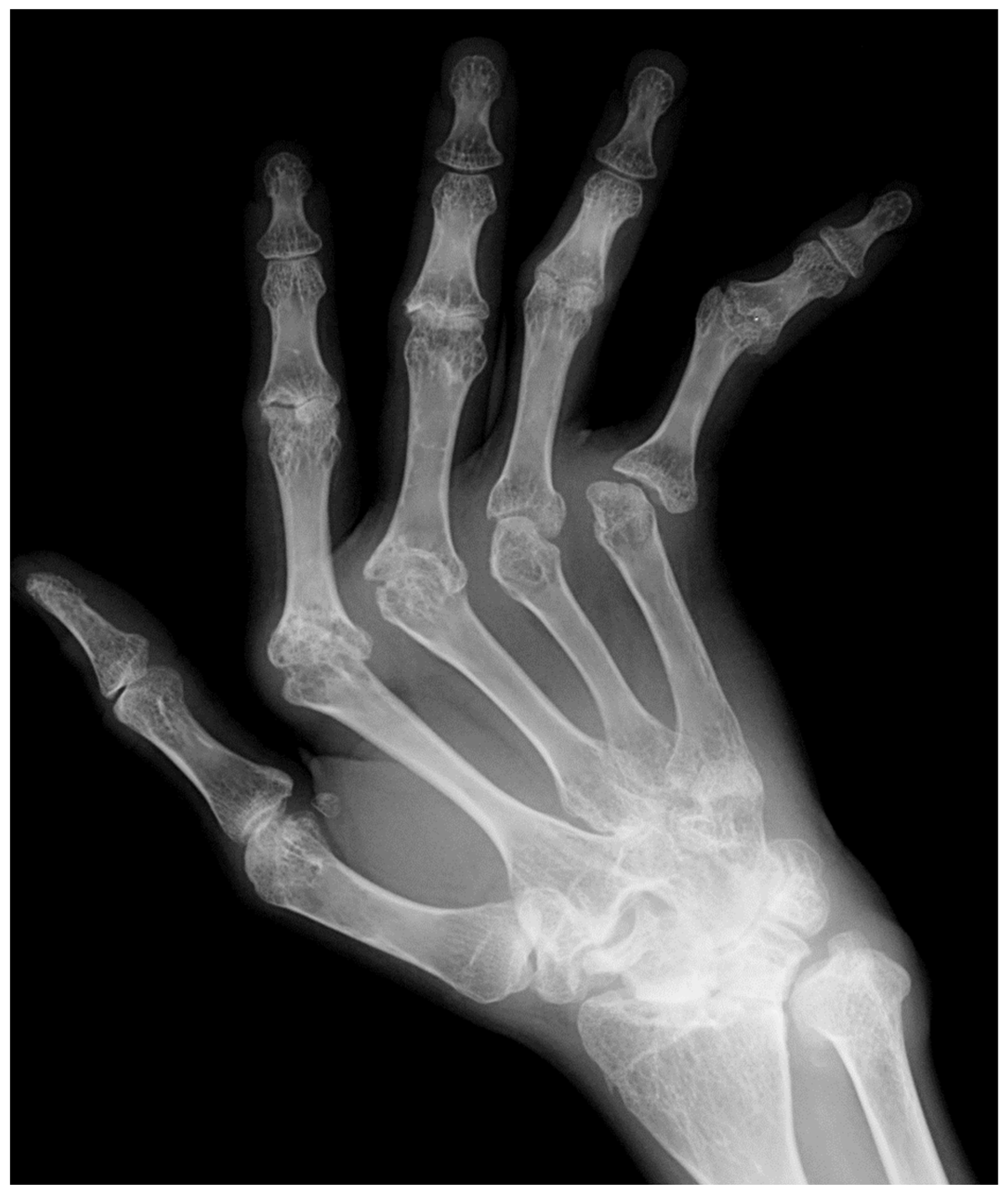
Rheumatoid Arthritis вђ Hand Radiology At St Vincent S University Introduction. rheumatoid arthritis (ra) is a symmetric, inflammatory, peripheral polyarthritis of unknown etiology. it typically leads to joint destruction through the erosion of cartilage and bone. untreated, it will lead to loss of physical function, inability to carry out daily tasks of living, and difficulties in maintaining employment. X ray images can help diagnose rheumatoid arthritis by showing changes in your bones and joints. ezzati f, et al. (2022). radiographic findings of inflammatory arthritis and mimics in the.

Rheumatoid Arthritis X Ray Foot

Comments are closed.