Oral Cavity Anatomy Mucosa My Xxx Hot Girl

Oral Cavity Anatomy Mucosa My Xxx Hot Girl Oral cavity. the oral cavity is situated anteriorly on the face, under the nasal cavities.it is bounded by a roof, a floor and lateral walls. anteriorly it opens to the face through the oral fissure, while posteriorly the oral cavity communicates with the oropharynx through a narrow passage called the oropharyngeal isthmus (also termed the isthmus of the fauces). Classification of oral mucosa. oral mucosa almost continuously lines the oral cavity. oral mucosa is composed of stratified squamous epithelium overlying a connective tissue proper, or lamina propria, with possibly a deeper submucosa (figure 9 1; see chapter 8). figure 9 1 general histological features of an oral mucosa composed of stratified.

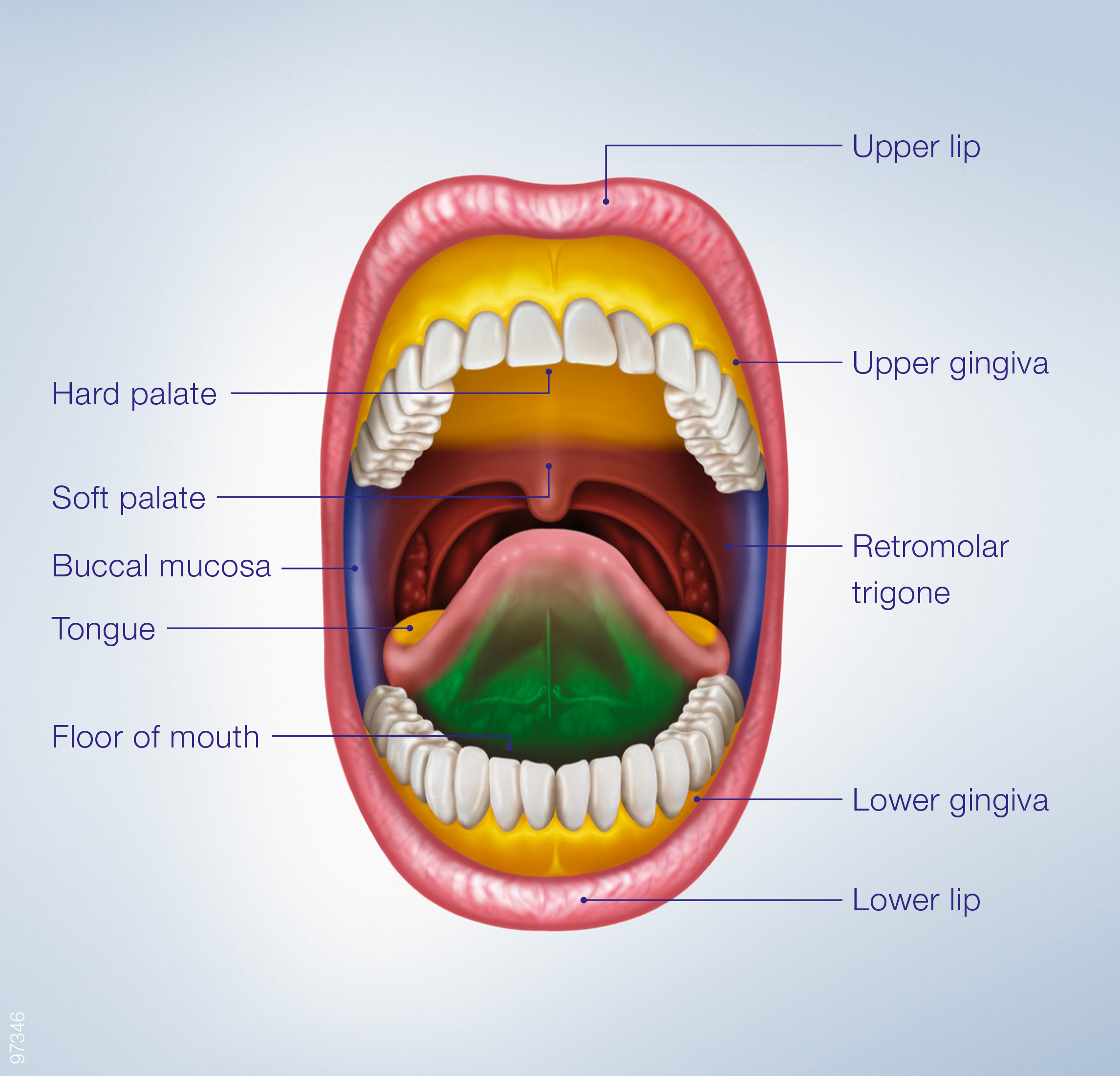
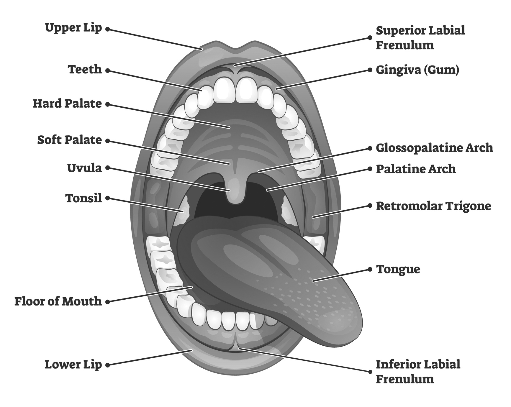
Oral Cavity Anatomy Mucosa My Xxx Hot Girl Vrogue Co Anatomy of the oral cavity. figure 1. anterior view of the a external mouth and lips and b arterial supply to the lips. figure 2. inferior view of the maxilla. figure 3. cross section of a tooth. figure 4. lateral cross section showing the a innervation of the lips b and teeth and gingiva. The oral cavity, better known as the mouth, is the start of the alimentary canal. it has three major functions: digestion – receives food, preparing it for digestion in the stomach and small intestine. communication – modifies the sound produced in the larynx to create a range of sounds. breathing – acts as an air inlet in addition to the. The oral cavity, or more commonly known as the mouth or buccal cavity, serves as the first portion of the digestive system. it consists of several different anatomically different aspects that work together effectively and efficiently to perform several functions. these aspects include the lips, tongue, palate, and teeth. although a small compartment, the oral cavity is a unique and complex. The oral mucosa forms a protective lining on the internal surface of the oral cavity. it extends from the labial borders to the pharyngeal mucosa posteriorly. it’s divided into three general types, including the lining mucosa, masticatory mucosa, and specialized mucosa. each of these three regions have different functions in mastication.

Oral Cavity Definition Anatomy Functions Diagram My Xxx Hot G The oral cavity, or more commonly known as the mouth or buccal cavity, serves as the first portion of the digestive system. it consists of several different anatomically different aspects that work together effectively and efficiently to perform several functions. these aspects include the lips, tongue, palate, and teeth. although a small compartment, the oral cavity is a unique and complex. The oral mucosa forms a protective lining on the internal surface of the oral cavity. it extends from the labial borders to the pharyngeal mucosa posteriorly. it’s divided into three general types, including the lining mucosa, masticatory mucosa, and specialized mucosa. each of these three regions have different functions in mastication. 5. attached gingiva. 6. alveolar mucosa. 7. labial frenum. at the margin or edge of the tooth, light pink, not attached to underlying structures, forms the soft wall of the gingival sulcus, perio probe is inserted under it, it is the first tissue to respond to inflammation or gingivitis. marginal or free gingiva. We have created 114 medical original illustrations of the mouth, the buccal cavity, the bones of the palate, the tongue, the salivary glands and the oral part of the pharynx with vessels and nerves. the drawing and anatomical labeling (using the terminologia anatomica 2) of these illustrations was done by gauthier kervyn under the anatomical.

Oral Cavity Anatomy Mucosa My Xxx Hot Girl Vrogue Co 5. attached gingiva. 6. alveolar mucosa. 7. labial frenum. at the margin or edge of the tooth, light pink, not attached to underlying structures, forms the soft wall of the gingival sulcus, perio probe is inserted under it, it is the first tissue to respond to inflammation or gingivitis. marginal or free gingiva. We have created 114 medical original illustrations of the mouth, the buccal cavity, the bones of the palate, the tongue, the salivary glands and the oral part of the pharynx with vessels and nerves. the drawing and anatomical labeling (using the terminologia anatomica 2) of these illustrations was done by gauthier kervyn under the anatomical.

Oral Cavity Anatomy Mucosa My Xxx Hot Girl Vrogue Co

Comments are closed.