Only Toppers Dare To Study Pressure Volume Diagram During The Cardiac

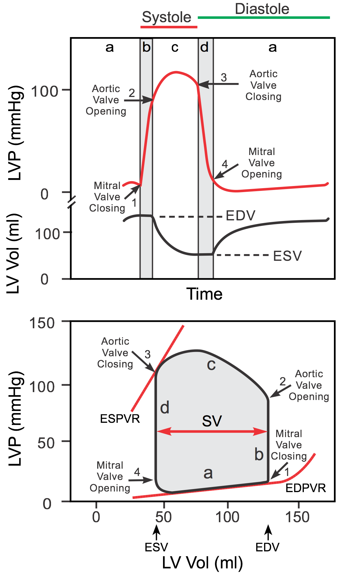
Only Toppers Dare To Study Pressure Volume Diagram During The Cardiac Find notes here: nonstopneuron post pressure volume diagram during the cardiac cycleexplore our entire animation video library: n. Ventricular pressure volume relationship. left ventricular pressure volume relationship can be described by a loop diagram with volume depicted on the x axis and left ventricular pressure on the y axis. if left ventricular pressure and volume are measured continuously during a single cardiac cycle, the loop diagram shown in figure 1 is obtained.

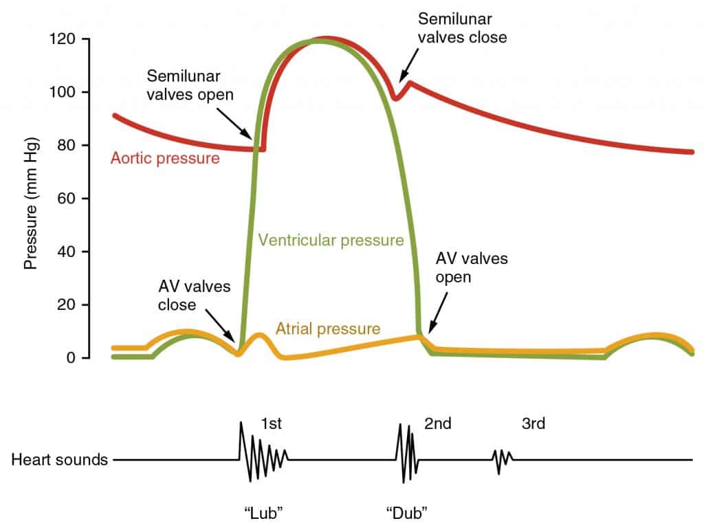
Cardiac Pressure Volume Loop1316278143849 Jpg 774г 702 Cardiology Left ventricular pressure volume (pv) loops are derived from pressure and volume information found in the cardiac cycle diagram (upper panel of the figure). to generate a pv loop for the left ventricle, the left ventricular pressure (lvp) is plotted against the left ventricular volume (lv vol) at multiple time points during a complete cardiac. Pressure volume (pv) analysis addresses these shortcomings by combining simultaneous measurements of pressure and volume to generate load independent measures of systolic and diastolic chamber properties, and is considered the gold standard method for characterizing ventricular systolic and diastolic function, as well as ventricular vascular. Wigger’s diagram is used to demonstrate the varying pressures in the atrium, ventricle, and artery during one cardiac cycle (figure 2). intracardiac pressures are different within the right and left sides of the heart. the left side has higher pressure, as it has to pump blood through the whole body, compared to the right side, which has to. Blood flow throughout the heart each cardiac cycle presents a complex pressure volume relationship. this relationship is comprised of various stages such as; diastole, isovolumetric contraction, systole, and isovolumetric relaxation. edv represents the blood volume found in the left ventricle after diastole, where the heart undergoes relaxation.

The Cardiac Cycle Describe The Changes Of Pressure And Volume That Wigger’s diagram is used to demonstrate the varying pressures in the atrium, ventricle, and artery during one cardiac cycle (figure 2). intracardiac pressures are different within the right and left sides of the heart. the left side has higher pressure, as it has to pump blood through the whole body, compared to the right side, which has to. Blood flow throughout the heart each cardiac cycle presents a complex pressure volume relationship. this relationship is comprised of various stages such as; diastole, isovolumetric contraction, systole, and isovolumetric relaxation. edv represents the blood volume found in the left ventricle after diastole, where the heart undergoes relaxation. The cardiac cycle is defined as a sequence of alternating contraction and relaxation of the atria and ventricles in order to pump blood throughout the body. it starts at the beginning of one heartbeat and ends at the beginning of another. the process begins as early as the 4th gestational week when the heart first begins contracting. A pv loop plots the changes in ventricular pressure associated with the changes in volume which occur during the cardiac cycle. multiple pieces of information can be extracted from such a loop, including volumes (eg. stroke volume), pressures (eg. end systolic pressure), and area (which represents myocardial work). the pv loop can also be used to represent the relationships of contractility.

Pressure And Volume Changes During Cardiac Cycle At Brenda Smith Blog The cardiac cycle is defined as a sequence of alternating contraction and relaxation of the atria and ventricles in order to pump blood throughout the body. it starts at the beginning of one heartbeat and ends at the beginning of another. the process begins as early as the 4th gestational week when the heart first begins contracting. A pv loop plots the changes in ventricular pressure associated with the changes in volume which occur during the cardiac cycle. multiple pieces of information can be extracted from such a loop, including volumes (eg. stroke volume), pressures (eg. end systolic pressure), and area (which represents myocardial work). the pv loop can also be used to represent the relationships of contractility.

Pressure And Volume Changes During Cardiac Cycle At Brenda Smith Blog

Comments are closed.