Nuclear Stress Myocardial Perfusion Imaging University Of Ottawa

Nuclear Stress Myocardial Perfusion Imaging University Of Ottawa Please arrive 15 minutes early. for more information, see coming for a diagnostic test. the nuclear stress test takes about four hours to complete. if you have any questions prior to your nuclear stress test, please call 613 696 7000 x14934, monday to friday, 7:00 a.m. to 4:00 p.m. Cardiac imaging. cardiac imaging enables physicians to rule out or validate evidence of coronary artery disease and provide early, effective treatments for patients. a variety of imaging methods are used at the heart institute. each has advantages and helps physicians understand how best to treat aspects of heart disease.

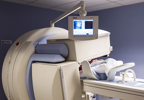
Stress Myocardial Perfusion Imaging University Of Ottawa Heart I Nuclear cardiology . nuclear stress myocardial perfusion imaging (mpi) is a nuclear cardiology test that shows how well blood flows to the muscle of the heart (myocardium). this test is used to diagnose the presence or absence of coronary artery disease. for more information about this test including what to expect and how to prepare for your. Directions and parking. general campus 501 smyth road main level tel.: 613 761 4831, option 8 hours: mon. – fri., 7:00 a.m. – 4:00 p.m. directions: from the main entrance, follow the signs on the main level (located at the public elevators). patients may also ask for directions at the patient information desk. Nuclear myocardial perfusion imaging (mpi) includes single photon emission computed tomography (spect) and positron emission tomography (pet). both tests enable relative perfusion imaging to identify ischemia in the myocardium, and gated imaging, which assesses wall motion abnormalities. unique to pet is the assessment of myocardial blood flow. Last reviewed: aug 18, 2023. myocardial perfusion imaging (mpi) is a non invasive imaging test that shows how well blood flows through your heart muscle. it can show areas of the heart muscle that aren’t getting enough blood flow. it can also show how well the heart muscle is pumping. this test is often called a nuclear stress test.

Nuclear Stress Myocardial Perfusion Imaging University Of Ottawa Nuclear myocardial perfusion imaging (mpi) includes single photon emission computed tomography (spect) and positron emission tomography (pet). both tests enable relative perfusion imaging to identify ischemia in the myocardium, and gated imaging, which assesses wall motion abnormalities. unique to pet is the assessment of myocardial blood flow. Last reviewed: aug 18, 2023. myocardial perfusion imaging (mpi) is a non invasive imaging test that shows how well blood flows through your heart muscle. it can show areas of the heart muscle that aren’t getting enough blood flow. it can also show how well the heart muscle is pumping. this test is often called a nuclear stress test. Cardiac stress imaging can assess coronary perfusion, cardiac (including valvular) function, myocardium viability, and exercise capacity.[1] stress can be induced pharmacologically or through physical exercise. imaging modalities include echocardiography and nuclear myocardial perfusion imaging with either single photon emission computed tomography (spect) or positron emission tomography (pet. Affiliations. 1 division of cardiovascular medicine and frankel cardiovascular center, university of michigan, ann arbor, michigan [email protected]. 2 mid america heart institute and the saint luke's health system, university of missouri kansas city, kansas city, missouri. 3 department of diagnostic radiology and nuclear medicine.

Comments are closed.