Myringotomy And Grommet Insertion Download Scientific Diagram
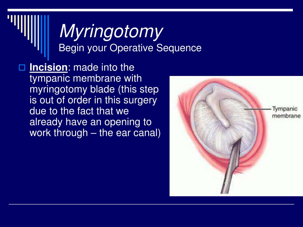
Myringotomy And Grommet Insertion Download Scientific Diagram Download scientific diagram | myringotomy and grommet insertion. from publication: how i do it: a simulator of the ear for developing otomicroscopy skills during the coronavirus disease 2019. Alternatively, emla cream® (lidocaine 2.5% and prilocaine 2.5%) can be applied to the tympanic membrane 30 minutes prior to the procedure, or the deep ear canal may be injected with local anesthesia with a dental needle. figure 1: ear speculum in place right ear with radial incision placed anteroinferiorly. figure 2: typical myringotomy knife.

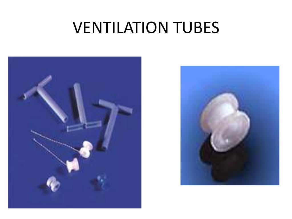
Myringotomy Grommet Tube Insertion Surgical – myringotomy and grommet insertion current guidance recommends the insertion of grommets * for those with >3 months of bilateral ome and hearing level in better ear > 25 30db hl. in certain cases, if there are significant concerns with speech and language development, grommets can be considered even if the hearing thresholds were. What is a myringotomy and ventilation tube insertion? a myringotomy is a small cut in the tympanic membrane (ear drum). a small tube (a grommet or t tube) is placed in this cut to allow for ventilation of the middle ear. grommets tend to remain in place for 6–18 months. t tubes are designed to be long term. Download scientific diagram | politzer's grommet. (reproduced with permission.) 31 from publication: history of myringotomy and grommets | the first recorded myringotomy was in 1649. astley cooper. Myringotomy is a simple procedure to drain a build up of fluid in the middle ear. a small incision is made in the ear drum (tympanic membrane) and the fluid is allowed to drain or is suctioned clear. often, tiny, self retaining plastic ear tubes (sometime called grommets) are inserted into the eardrum. the operation, which may take up to half.

Myringotomy And Grommet Insertion Download scientific diagram | politzer's grommet. (reproduced with permission.) 31 from publication: history of myringotomy and grommets | the first recorded myringotomy was in 1649. astley cooper. Myringotomy is a simple procedure to drain a build up of fluid in the middle ear. a small incision is made in the ear drum (tympanic membrane) and the fluid is allowed to drain or is suctioned clear. often, tiny, self retaining plastic ear tubes (sometime called grommets) are inserted into the eardrum. the operation, which may take up to half. Tiny tubes called grommets are then inserted into these cuts. the main function of a grommet is to act as a ventilation tube – the grommet lets air pass from the ear tube through the eardrum and into the middle ear. any fluid in the middle ear will now just dry up. you should be able to return to normal activity after 24 48 hours. Indication of when and how to perform a paracentesis (cutting the eardrum) and how to insert a ventilation tube into the tympanic membrane is explained in de.

Ppt Myringotomy With Ear Tubes Powerpoint Presentation Free Download Tiny tubes called grommets are then inserted into these cuts. the main function of a grommet is to act as a ventilation tube – the grommet lets air pass from the ear tube through the eardrum and into the middle ear. any fluid in the middle ear will now just dry up. you should be able to return to normal activity after 24 48 hours. Indication of when and how to perform a paracentesis (cutting the eardrum) and how to insert a ventilation tube into the tympanic membrane is explained in de.

Comments are closed.