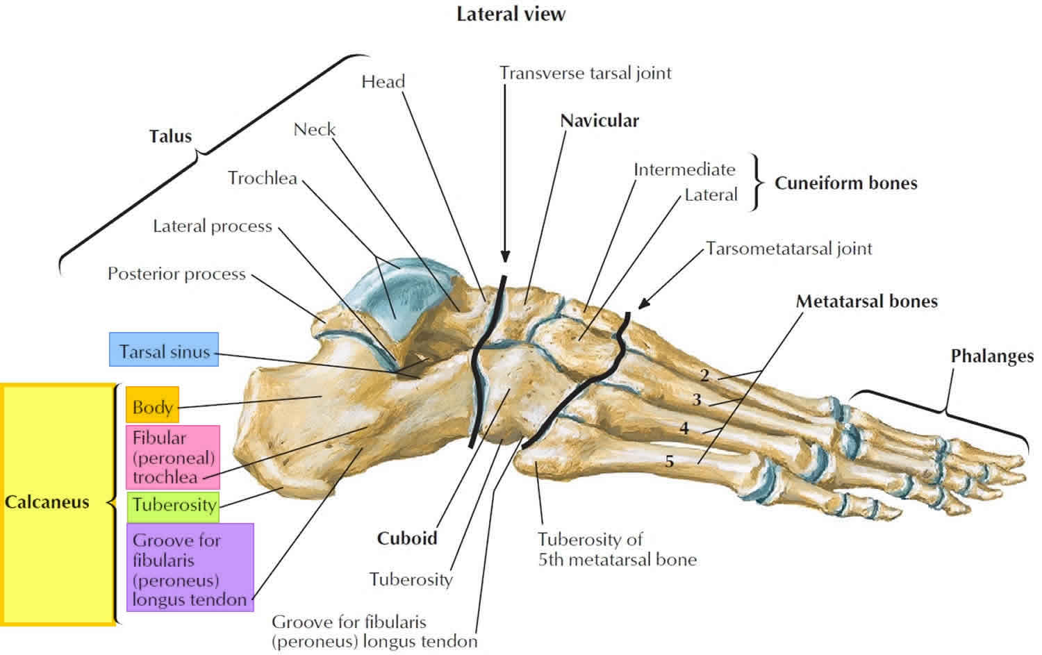
Lateral View Of The Osteology Of The Foot Ankle Complex Several Bones

Lateral View Of The Osteology Of The Foot Ankle Complex Several Bones Download scientific diagram | lateral view of the osteology of the foot ankle complex. several bones including the medial cuneiform, first metatarsal, and first phalanges are not shown. from. The kinematics of the ankle and foot may be the most complex in the human body. many irregularly shaped joints are capable of producing unique motions not yet introduced in this text. two sets of terminology are therefore necessary to fully describe the complex kinematics at the ankle and foot: fundamental and applied.

The Ligament Configuration Of The Foot Lateral View Picture The foot and the ankle work synchronically in a complex system made of 26 bones and joints. they help the feet to adapt to uneven surfaces while the other parts of the body rest standing. the ankle plays an essential role in the execution of plantar flexion and dorsiflexion movements. the foot can be subdivided into the hindfoot, the midfoot. Ankle anatomy. the ankle joint, also known as the talocrural joint, allows dorsiflexion and plantar flexion of the foot. it is made up of three joints: upper ankle joint (tibiotarsal), talocalcaneonavicular, and subtalar joints. the last two together are called the lower ankle joint. The role of the foot and ankle is multifaceted, providing a supportive base capable of withstanding body loading while simultaneously permitting mobility. this chapter provides an overview of the bony anatomy of the foot and ankle and its complex articulations, muscle, and ligamentous attachments. download reference work entry pdf. Introduction. this topic introduces the osteology (the structure and function) of the bones of the foot and ankle. including the: calceneous bones. lateral malleous. sinus tarsi. talar dome, and. talonavicular joint. cricos no. 00213j | abn 83791 724 622.

Calcaneus Bone Anatomy Function Calcaneus Pain Calcaneus Fracture The role of the foot and ankle is multifaceted, providing a supportive base capable of withstanding body loading while simultaneously permitting mobility. this chapter provides an overview of the bony anatomy of the foot and ankle and its complex articulations, muscle, and ligamentous attachments. download reference work entry pdf. Introduction. this topic introduces the osteology (the structure and function) of the bones of the foot and ankle. including the: calceneous bones. lateral malleous. sinus tarsi. talar dome, and. talonavicular joint. cricos no. 00213j | abn 83791 724 622. Anatomy. saddle shaped. ligament support. plantar support is by the superficial and deep inferior calcaneocuboid ligaments. superior support is by the lateral limb of the bifurcate ligamant. motion. inversion of subtalar joint locks the transverse tarsal joint. allows for a stable hindfoot midfoot for toe off. The foot is the most complex structure, formed by 28 bones, 33 joints, and 112 ligaments, which are controlled by extrinsic and intrinsic muscles. the foot can be divided into the hindfoot (talus and calcaneus bones); the midfoot, which is formed by the cuboid, navicular, and three cuneiform bones; and the forefoot.

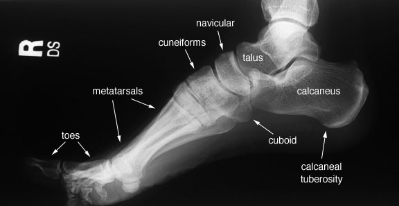
Radiographic Anatomy Of The Skeleton Foot Lateral View Labelled Anatomy. saddle shaped. ligament support. plantar support is by the superficial and deep inferior calcaneocuboid ligaments. superior support is by the lateral limb of the bifurcate ligamant. motion. inversion of subtalar joint locks the transverse tarsal joint. allows for a stable hindfoot midfoot for toe off. The foot is the most complex structure, formed by 28 bones, 33 joints, and 112 ligaments, which are controlled by extrinsic and intrinsic muscles. the foot can be divided into the hindfoot (talus and calcaneus bones); the midfoot, which is formed by the cuboid, navicular, and three cuneiform bones; and the forefoot.

Lateral Aspect Of The Ankle Ligaments Netter Ankle Anatomy Foot

Comments are closed.