Labeled Diagram Of A Kidney

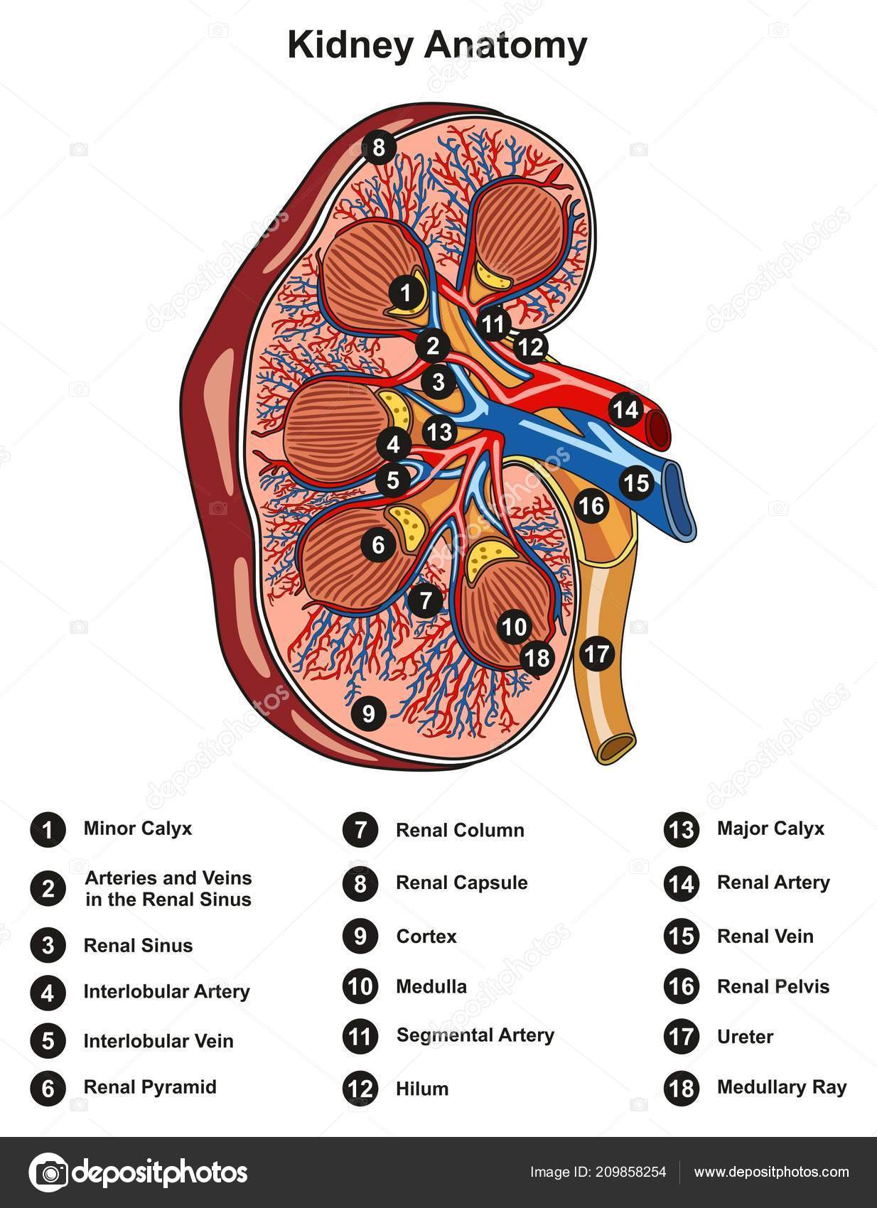
Diagram Of A Kidney Labeled Internal anatomy of the kidney (overview) the main unit of the medulla is the renal pyramid. there are 8 18 renal pyramids in each kidney, that on the coronal section look like triangles lined next to each other with their bases directed toward the cortex and apex to the hilum. the apex of the pyramid projects medially toward the renal sinus. The kidneys are two bean shaped organs in the renal system. they help the body pass waste as urine. they also help filter blood before sending it back to the heart. the kidneys perform many.

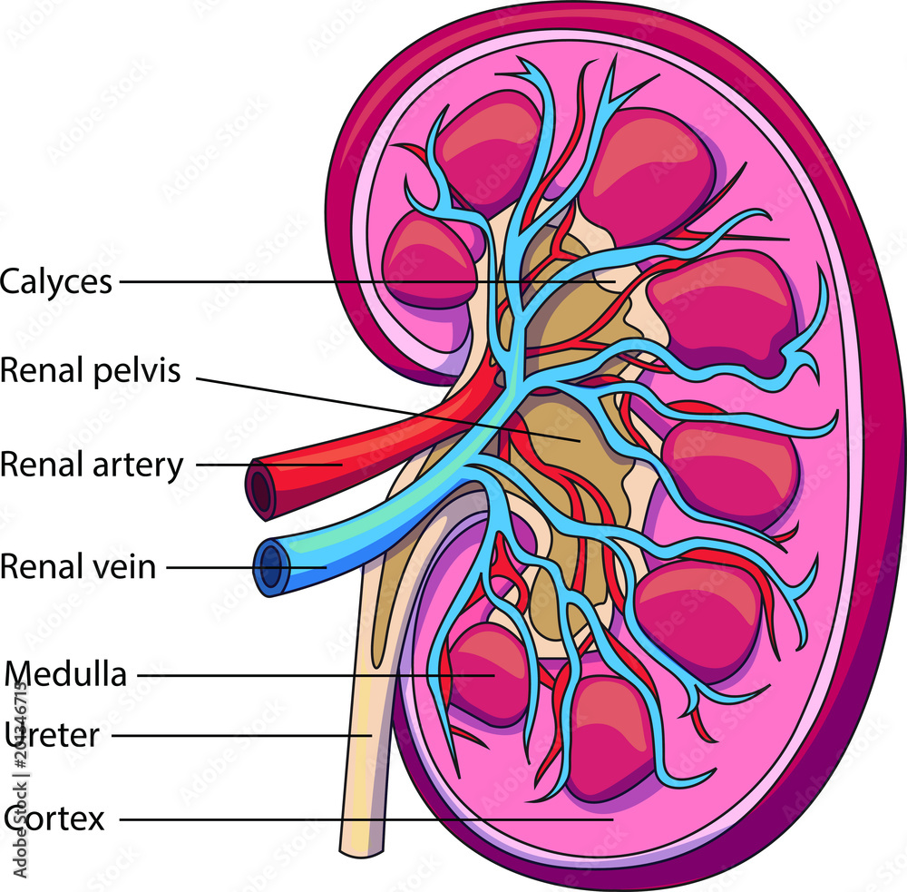
Biology Mbbs Structure Of Human Kidney With Labeled Diagram Ratta Pk The kidneys. the kidneys are bilateral bean shaped organs, reddish brown in colour and located in the posterior abdomen. their main function is to filter and excrete waste products from the blood. they are also responsible for water and electrolyte balance in the body. metabolic waste and excess electrolytes are excreted by the kidneys to form. The kidneys. explore the anatomy, structure, and role of the kidneys with innerbody's interactive 3d model. the kidneys are the waste filtering and disposal system of the body. as much as 1 3 of all blood leaving the heart passes into the kidneys to be filtered before flowing to the rest of the body's tissues. Acute renal failure or acute kidney injury occurs quickly, with fluids and waste products building up and causing a cascade of problems in the body. causes include toxins, shock, sepsis, cardiac issues, and more. chronic kidney disease: this is the result of long term kidney damage that gradually reduces the function of the kidneys. while some. Kidney anatomy. renal capsule – an outer membrane that surrounds the kidney; it is thin but tough and fibrous. renal pelvis – basin like area that collects urine from the nephrons (the kidney’s filtration system), it narrows into the upper end of the ureter. calyx – the extension of the renal pelvis; they channel urine from the pyramids.

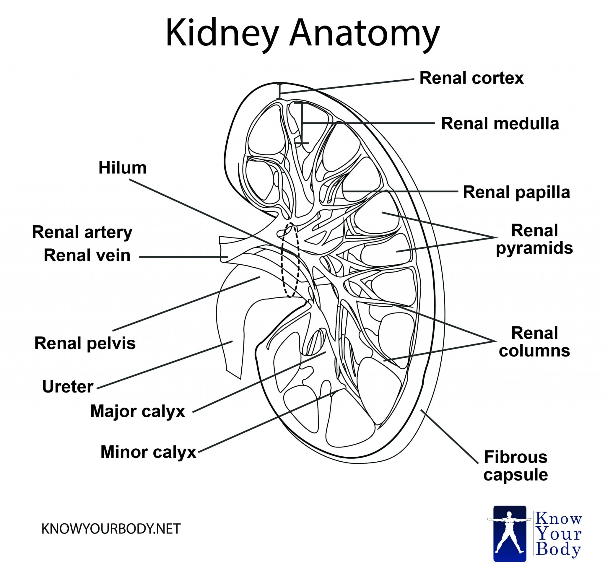
Kidney Location Function Anatomy Diagram And Faqs Acute renal failure or acute kidney injury occurs quickly, with fluids and waste products building up and causing a cascade of problems in the body. causes include toxins, shock, sepsis, cardiac issues, and more. chronic kidney disease: this is the result of long term kidney damage that gradually reduces the function of the kidneys. while some. Kidney anatomy. renal capsule – an outer membrane that surrounds the kidney; it is thin but tough and fibrous. renal pelvis – basin like area that collects urine from the nephrons (the kidney’s filtration system), it narrows into the upper end of the ureter. calyx – the extension of the renal pelvis; they channel urine from the pyramids. Internal anatomy. a frontal section through the kidney reveals an outer region called the renal cortex and an inner region called the renal medulla (figure 25.1.2). in the medulla, 5 8 renal pyramids are separated by connective tissue renal columns. each pyramid creates urine and terminates into a renal papilla. Kidney anatomy. the shape of each kidney gives it a convex side and a concave side. you can see this clearly in the detailed diagram of kidney anatomy shown in figure \(\pageindex{3}\). the concave side is where the renal artery enters the kidney and the renal vein and ureter leave the kidney. this area of the kidney is called the hilum.

Schematic Vector Diagram Of A Kidney Kidney Structure With Labeled Internal anatomy. a frontal section through the kidney reveals an outer region called the renal cortex and an inner region called the renal medulla (figure 25.1.2). in the medulla, 5 8 renal pyramids are separated by connective tissue renal columns. each pyramid creates urine and terminates into a renal papilla. Kidney anatomy. the shape of each kidney gives it a convex side and a concave side. you can see this clearly in the detailed diagram of kidney anatomy shown in figure \(\pageindex{3}\). the concave side is where the renal artery enters the kidney and the renal vein and ureter leave the kidney. this area of the kidney is called the hilum.

Comments are closed.