Introduction To Abdomen Radiography Radiology Key

Introduction To Abdomen Radiography Radiology Key Limitations. a caveat to keep in mind is that the diagnostic yield of plain film of the abdomen is low in the absence of signs and symptoms; therefore, an abdomen radiograph usually is not recommended as a survey tool. box 28 1 lists many conditions for which plain film radiographs are not indicated because roentgenographic findings rarely occur. Nearly all ct scans of the abdomen require the administration of intraluminal contrast agents to demonstrate the lumen of and to distend the gastrointestinal tract. radiopaque agents may be dilute concentrations of barium or iodinated contrast agents. iodinated agent concentrations of 1% to 3% are optimal for intraluminal opacification for ct.

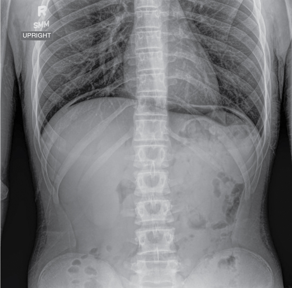
Abdominal Radiography Radiology Key Abdominal radiographs may be obtained in the radiology department or may be performed portably. views should generally include either the diaphragm or inferior pubic ramus. gonadal shielding may be provided for men. portable abdominal radiographs may be necessary due to patient immobility but are of much poorer quality. Study with quizlet and memorize flashcards containing terms like who is qualified to verify a first or second competency when a student performs a procedure? select one: a. radiologist b. radiographer c. radiology manager, the main concern at clinical for student medical radiographers is: select one: a. working with the radiologist in a professional manner b. treating the patient with respect. Introduction to computed tomographic imaging of the chest. abdominal anatomy on computed tomography. normal renal anatomy. introduction to musculoskeletal radiology. introduction to spine radiographs. introduction to brain surface anatomy. abdominal x rays. 1 introduction to abdominal radiography. ajadi adetola department of veterinary medicine and surgery. 2 abdominal radiography: indications. vomiting abdominal pain regurgitation abdominal masses diarrhea haematuria dysuria tenesmus herniation rectal bleeding suspected foreign body staging of neoplasia others. 6 abdominal radiography: technical.

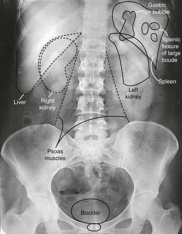
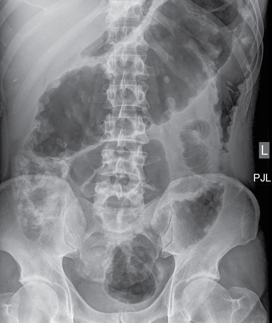
Abdominal Radiography Radiology Key Vrogue Co Introduction to computed tomographic imaging of the chest. abdominal anatomy on computed tomography. normal renal anatomy. introduction to musculoskeletal radiology. introduction to spine radiographs. introduction to brain surface anatomy. abdominal x rays. 1 introduction to abdominal radiography. ajadi adetola department of veterinary medicine and surgery. 2 abdominal radiography: indications. vomiting abdominal pain regurgitation abdominal masses diarrhea haematuria dysuria tenesmus herniation rectal bleeding suspected foreign body staging of neoplasia others. 6 abdominal radiography: technical. A systematic approach to abdominal x ray interpretation is therefore relatively straightforward. this involves assessment of the bowel gas pattern, soft tissue structures, and bones. full assessment includes a check of patient data, image quality, and checking for artifact and abnormal calcification. this tutorial will discuss these steps. Mammography is an x‐ray‐based imaging modality that uses low‐energy x‐rays to image the breasts as a diagnostic and screening tool. image production. x‐ray tubes in mammography units used molybdenum as a target and a much smaller focal spots. the tube voltage in mammography ranges from 25 to 34 kv.

Abdominal Radiography Radiology Key A systematic approach to abdominal x ray interpretation is therefore relatively straightforward. this involves assessment of the bowel gas pattern, soft tissue structures, and bones. full assessment includes a check of patient data, image quality, and checking for artifact and abnormal calcification. this tutorial will discuss these steps. Mammography is an x‐ray‐based imaging modality that uses low‐energy x‐rays to image the breasts as a diagnostic and screening tool. image production. x‐ray tubes in mammography units used molybdenum as a target and a much smaller focal spots. the tube voltage in mammography ranges from 25 to 34 kv.

Comments are closed.