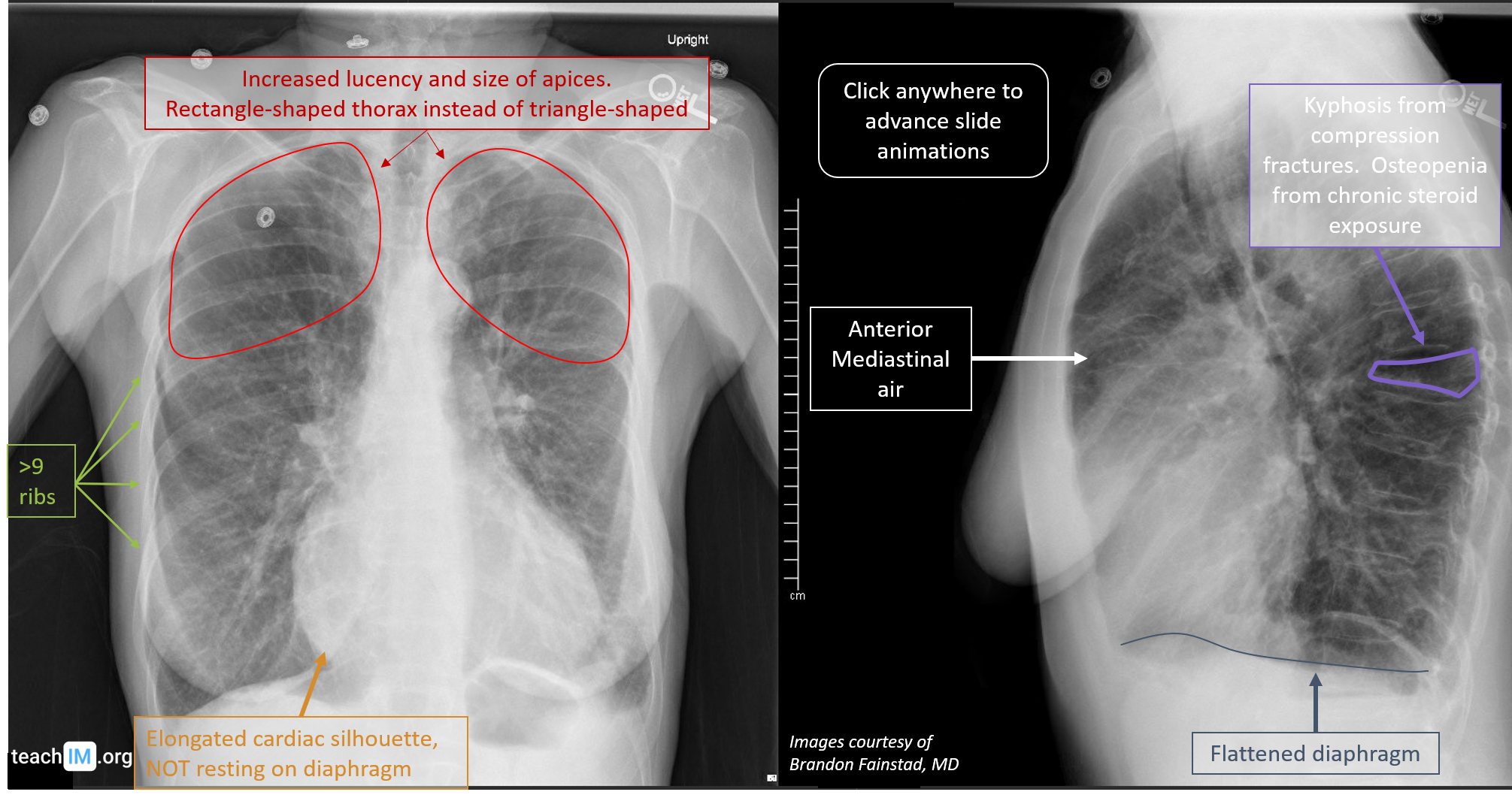
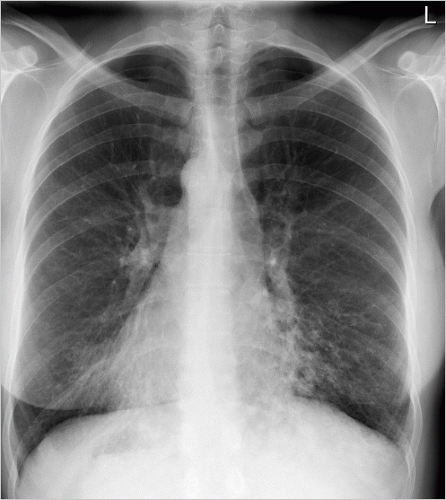
Imaging Of Chronic Obstructive Pulmonary Disease

Ct Imaging Of Chronic Obstructive Pulmonary Disease Insights The global initiative for chronic obstructive lung disease (gold) staging system is a commonly used severity staging system based on airflow limitation. according to this, there are four key stages with the latest revision at time of writing being in 2019 17: stage i: mild, fev 1 > 80% of normal. stage ii: moderate, fev 1 = 50 79% of normal. Chronic obstructive pulmonary disease (copd) is a complex disease with significant heterogeneity in terms of structural and clinical manifestations as well as progression. for instance, subjects with similar severity of airflow obstruction may experience marked differences in symptoms, health related quality of life, exacerbation prevalence, and exercise capacity.

Image Chronic Obstructive Pulmonary Disease Chest X Ray Msd Manual This article reviews the role of imaging in chronic obstructive pulmonary disease (copd), with a focus on common findings for the general radiologist. imaging features reviewed include visual and quantitative computed tomography assessment, nuclear medicine imaging, and mri. imaging advances and insights that have contributed to the genetic epidemiology of copd (copdgene) study in. Ct imaging is a readily quantifiable tool that can provide in vivo assessments of lung structure in conditions such as chronic obstructive pulmonary disease (copd). the information extracted from these data has been used in many clinical, epidemiological, and genetic investigations for patient stratification and prognostication, and to determine intermediate endpoints for clinical trials. Chronic obstructive pulmonary disease (copd) is a common and treatable disease characterized by progressive airflow limitation and tissue destruction. it is associated with structural lung changes due to chronic inflammation from prolonged exposure to noxious particles or gases most commonly cigarette smoke. chronic inflammation causes airway narrowing and decreased lung recoil. the disease. Lung imaging is increasingly being used to diagnose, quantify, and phenotype chronic obstructive pulmonary disease (copd). although spirometry is the gold standard for the diagnosis of copd and for severity staging, the role of computed tomography (ct) imaging has expanded in both clinical practice and research.

Chronic Obstructive Pulmonary Disease Copd Teachim Chronic obstructive pulmonary disease (copd) is a common and treatable disease characterized by progressive airflow limitation and tissue destruction. it is associated with structural lung changes due to chronic inflammation from prolonged exposure to noxious particles or gases most commonly cigarette smoke. chronic inflammation causes airway narrowing and decreased lung recoil. the disease. Lung imaging is increasingly being used to diagnose, quantify, and phenotype chronic obstructive pulmonary disease (copd). although spirometry is the gold standard for the diagnosis of copd and for severity staging, the role of computed tomography (ct) imaging has expanded in both clinical practice and research. The purpose of this statement is to describe and define the phenotypic abnormalities that can be identified on visual and quantitative evaluation of computed tomographic (ct) images in subjects with chronic obstructive pulmonary disease (copd), with the goal of contributing to a personalized approach to the treatment of patients with copd. Chronic obstructive pulmonary disease (copd) is characterized by the presence of airflow limitation that is caused by a combination of small airway remodeling and emphysema induced loss of elastic recoil. the management of copd depends on the relative distribution and severity of these two pathologic processes, factors that may vary widely even among patients with a similar degree of airflow.

Chronic Obstructive Pulmonary Disease Radiology Key The purpose of this statement is to describe and define the phenotypic abnormalities that can be identified on visual and quantitative evaluation of computed tomographic (ct) images in subjects with chronic obstructive pulmonary disease (copd), with the goal of contributing to a personalized approach to the treatment of patients with copd. Chronic obstructive pulmonary disease (copd) is characterized by the presence of airflow limitation that is caused by a combination of small airway remodeling and emphysema induced loss of elastic recoil. the management of copd depends on the relative distribution and severity of these two pathologic processes, factors that may vary widely even among patients with a similar degree of airflow.

Chronic Obstructive Pulmonary Disease Radiology Reference Article

Comments are closed.