Iii Radiology Lecture Abdominal And Git Radiology The Gastrointestinal Track

Iii Radiology Lecture Abdominal And Git Radiology The This is the 2020 edition of my talk on abdominal and git radiology. i have updated the talk since last year. Indications: i. dyspepsia ii. weight loss iii. upper abdominal mass iv. gastrointestinal haemorrhage or unexplained iron deficiency anaemia v. partial obstruction vi. assessment of site of perforation – it is essential that water soluble contrast medium e.g. gastrografin or dionosil aqueous is used. contraindications: i.

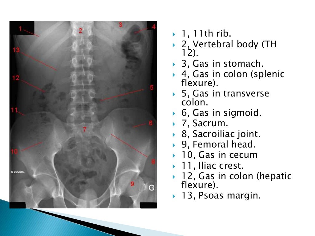
Radiographic Anatomy Of Gastrointestinal Tract Radiology lecture abdominal and git radiology the gastrointestinal track iii. radiology. The gastrointestinal tract may itself be subdivided into the upper and lower gastrointestinal tracts. the upper gastrointestinal tract usually refers to the structures from the mouth to the duodenum, whilst the lower gastrointestinal tract refers to all structures distal to the duodenojejunal flexure, i.e. small and large bowels to the anal verge. The gastrointestinal (gi) tract, also known as the alimentary canal, includes the lips, mouth, pharynx, esophagus, stomach, small intestine, appendix, and colon through to the anus. sonographic examination of the gi tract began in the 1980s using endoscopic ultrasonography (eus) and the transabdominal approach. Overview. the gastrointestinal (gi) tract, also known as the alimentary canal, commences at the buccal cavity of the mouth and terminates at the anus. it can be divided into an upper gi tract (consisting of mouth, pharynx, esophagus and stomach) and a lower gi tract (small and large intestines). the three primary functions of the gi tract are.

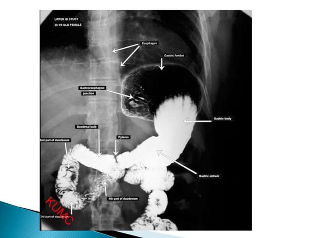
Radiographic Anatomy Of Gastrointestinal Tract The gastrointestinal (gi) tract, also known as the alimentary canal, includes the lips, mouth, pharynx, esophagus, stomach, small intestine, appendix, and colon through to the anus. sonographic examination of the gi tract began in the 1980s using endoscopic ultrasonography (eus) and the transabdominal approach. Overview. the gastrointestinal (gi) tract, also known as the alimentary canal, commences at the buccal cavity of the mouth and terminates at the anus. it can be divided into an upper gi tract (consisting of mouth, pharynx, esophagus and stomach) and a lower gi tract (small and large intestines). the three primary functions of the gi tract are. Esophageal barium swallow. 1 is a medical imaging procedure used to examine upper git, which include the esophagus. and to a lesser extent the stomach. 2 the contrast used is barium sulfate single contrast to assess anatomy or obstruction. double contrast to assess mucosal details. This section serves as an introduction to the basic anatomy of the abdomen. the human gastrointestinal tract can be divided into upper and lower portions. all structures proximal to the ligament of treitz can be thought of as the upper gi tract, while structures found distal to it are considered the lower gi tract.

Comments are closed.