Hepertensive Retinopathy Central Retinal Artery Occlusion Central

Hepertensive Retinopathy Central Retinal Artery Occlusion Central Moreover, hypertension can also cause occlusion of major retinal vessels such as the branch retinal artery, central retinal artery, branch retinal vein and central retinal vein. this article focuses primarily upon hypertensive retinopathy, which is the most common ocular presentation. disease entity disease. Hypertensive retinopathy is an eye condition in which high blood pressure damages the layer of tissue at the back of your eyeball (retina). high blood pressure means blood is pushing with more force than normal against your artery walls. over time, this pressure can damage your arteries and interfere with blood flow to various parts of your body.

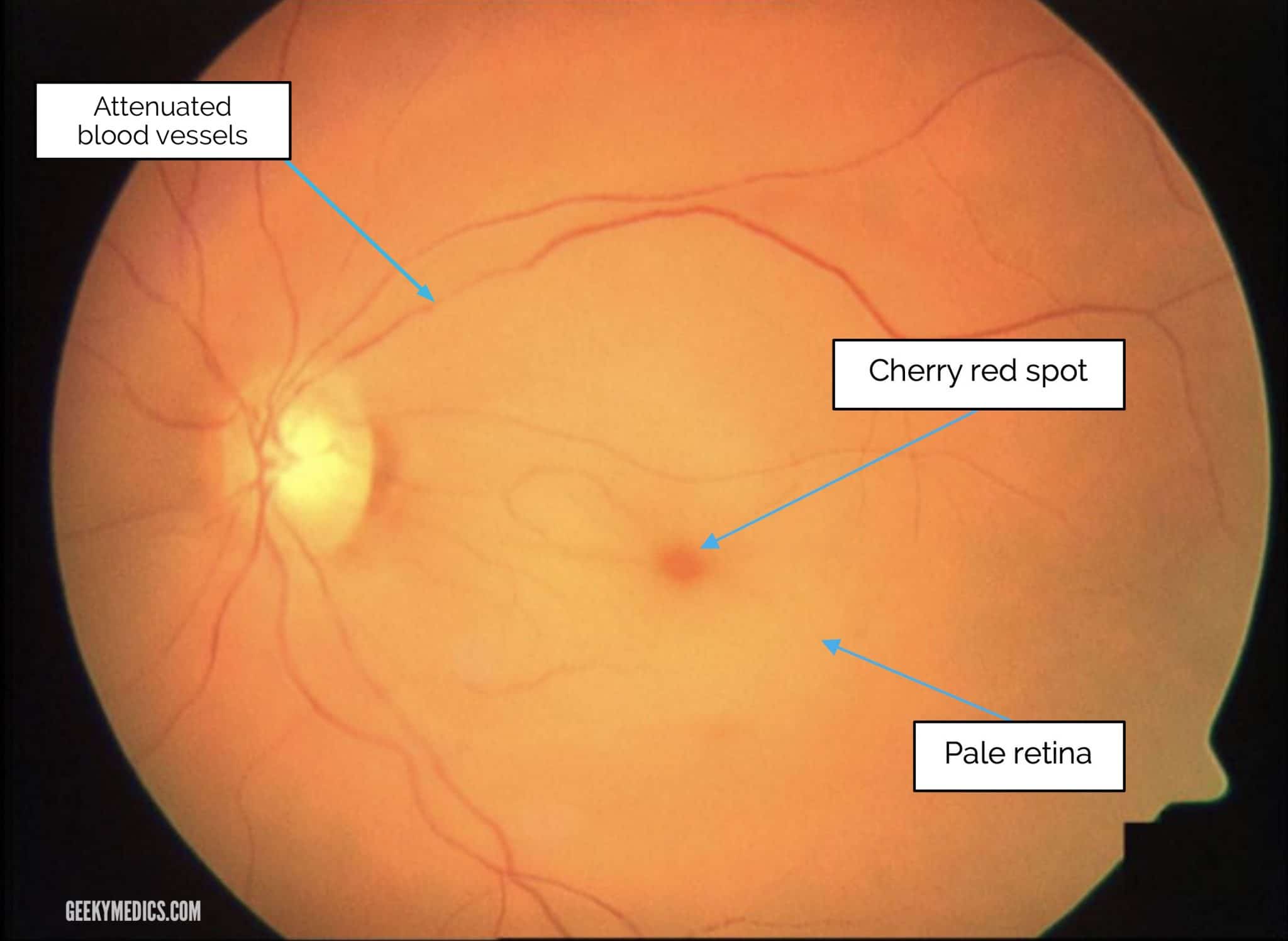
Central Retinal Artery Occlusion Crao Geeky Medics Central retinal artery occlusion (crao) is an ophthalmic emergency that can lead to sudden and severe vision loss.[1] crao has been defined as interruption of blood flow through the central retinal artery by thromboembolism or vasospasm with or without retinal ischemia. an embolus from the carotid artery, aortic arch, or heart often causes the condition. giant cell arteritis is another rare. Central retinal artery occlusion (crao) is an ocular emergency. patients typically present with profound, acute, painless monocular visual loss—with 80% of affected individuals having a final visual acuity of counting fingers or worse. crao is the ocular analogue of a cerebral stroke—and, as such, the clinical approach and management are. Central retinal artery occlusion (crao) is an ophthalmic emergency characterized by vision loss attributable to obstruction of blood flow through the central retinal artery (cra), 1 of the major arteries supplying blood to the eye. crao can be categorized into 4 distinct subtypes that can differ in cause, clinical presentation, and treatment. Poorly controlled hypertension (htn) affects several systems such as the cardiovascular, renal, cerebrovascular, and retina. the damage to these systems is known as target organ damage (tod).[1] htn affects the eye causing 3 types of ocular damage: choroidopathy, retinopathy, and optic neuropathy.[2] hypertensive retinopathy (hr) occurs when the retinal vessels get damaged due to elevated.

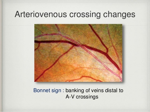
Hepertensive Retinopathy Central Retinal Artery Occlusion Central Central retinal artery occlusion (crao) is an ophthalmic emergency characterized by vision loss attributable to obstruction of blood flow through the central retinal artery (cra), 1 of the major arteries supplying blood to the eye. crao can be categorized into 4 distinct subtypes that can differ in cause, clinical presentation, and treatment. Poorly controlled hypertension (htn) affects several systems such as the cardiovascular, renal, cerebrovascular, and retina. the damage to these systems is known as target organ damage (tod).[1] htn affects the eye causing 3 types of ocular damage: choroidopathy, retinopathy, and optic neuropathy.[2] hypertensive retinopathy (hr) occurs when the retinal vessels get damaged due to elevated. This is called a central retinal artery occlusion (crao). your retina is the layer of nerve tissue at the back of your inner eye that senses light. the retina turns images into electrical signals. your optic nerve carries these signals to your brain. if a blockage of a blood vessel happens in your retina, it can be very serious. Hypertensive retinopathy is retinal vascular damage caused by hypertension. signs usually develop late in the disease. funduscopic examination shows arteriolar constriction, arteriovenous nicking, vascular wall changes, flame shaped hemorrhages, cotton wool spots, yellow hard exudates, and optic disk edema. treatment is directed at controlling.

Hepertensive Retinopathy Central Retinal Artery Occlusion Central This is called a central retinal artery occlusion (crao). your retina is the layer of nerve tissue at the back of your inner eye that senses light. the retina turns images into electrical signals. your optic nerve carries these signals to your brain. if a blockage of a blood vessel happens in your retina, it can be very serious. Hypertensive retinopathy is retinal vascular damage caused by hypertension. signs usually develop late in the disease. funduscopic examination shows arteriolar constriction, arteriovenous nicking, vascular wall changes, flame shaped hemorrhages, cotton wool spots, yellow hard exudates, and optic disk edema. treatment is directed at controlling.

Hepertensive Retinopathy Central Retinal Artery Occlusion Central

Comments are closed.