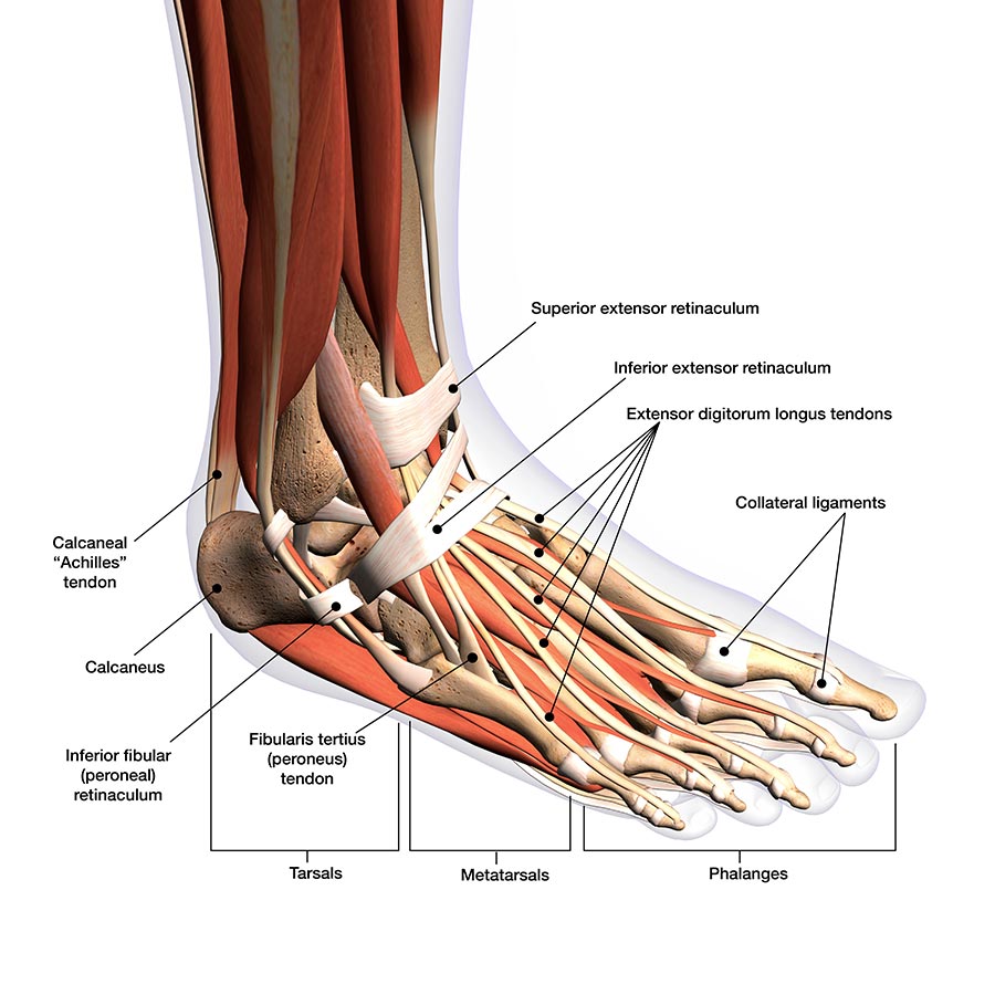
Foot And Ankle Anatomy Diagram

Foot Tendons Diagram Learn about the anatomy of the ankle and foot, including the bones, joints, ligaments, and muscles. see diagrams, videos, quizzes, and mnemonics to help you remember the key facts. Learn about the bones, joints, ligaments, muscles, tendons, and nerves of the foot and ankle. see diagrams and descriptions of the regions, columns, and essential joints of the foot.

Foot Anatomy Bones Joints Body Anatomy Learn about the ankle joint, a hinge type joint formed by the tibia, fibula and talus. see diagrams of the articulating surfaces, ligaments, muscles, and clinical relevance of ankle injuries. Dr. ebraheim’s educational animated video describes anatomical structures of the foot and ankle, the bony anatomy, the joints, ligaments, and the compartment. The foot and ankle form a complex system which consists of 28 bones, 33 joints, 112 ligaments, controlled by 13 extrinsic and 21 intrinsic muscles. the foot is subdivided into the rearfoot, midfoot, and forefoot. it functions as a rigid structure for weight bearing and it can also function as a flexible structure to conform to uneven terrain. Learn about the ankle joint, its structure, function, and common problems. see diagrams and videos of the bones, ligaments, tendons, muscles, nerves, and blood vessels that make up the ankle.

Comments are closed.