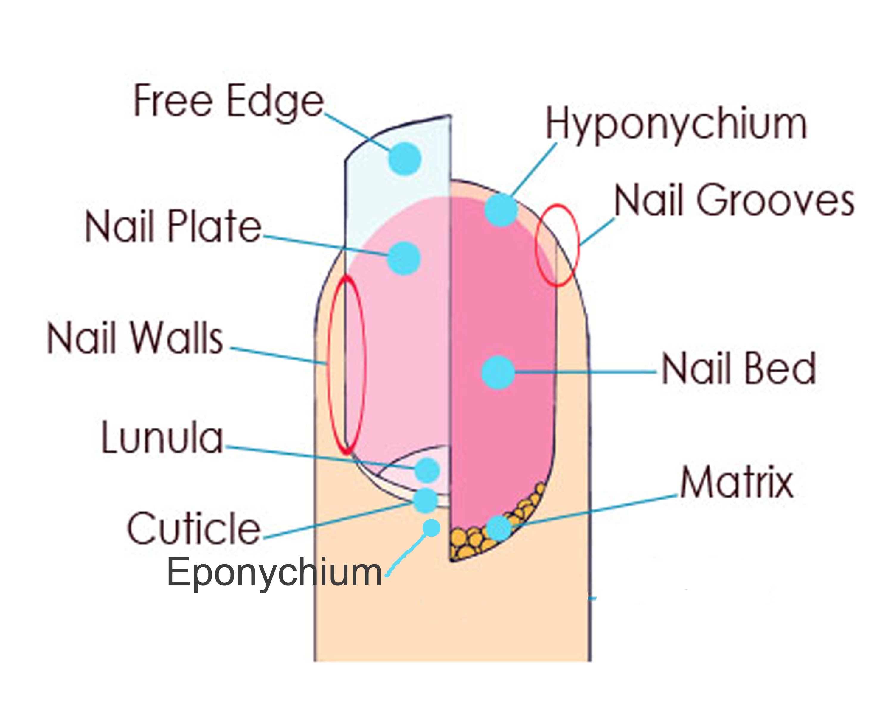
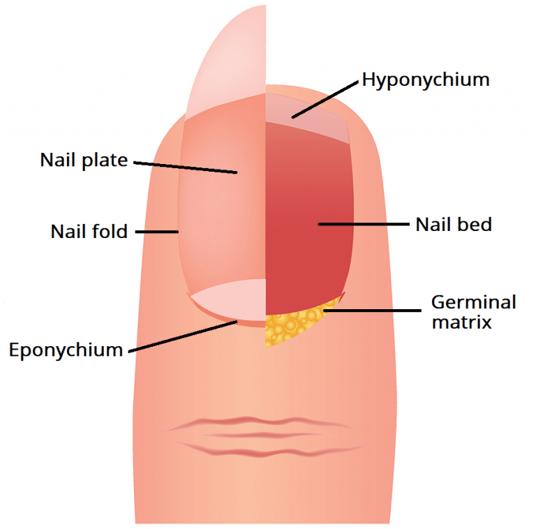
Diagram Of Nail Structure

Diagram Of The Nail And Its Structure Overview of nail anatomy. a nail is a flattish claw like plate at the tip of the fingers and toes, which is characteristically found in all primates. they are composed of the resilient protein keratin. nails are continuously growing and adjusting to their surroundings. they provide an essential protective shield for our fingertips. It produces most of the volume of the nail and the nail bed. the germinal matrix (the nail matrix) and the nail root are related. the matrix lies beneath the skin, at the inner edge of the nail plate, and is responsible for most of a nail's growth. it's where new cells grow and then advance forward to form the nail, until it reaches the outer.

Diagram Of The Nail And Its Structure Learn about the structure and function of the nail bed, nail body, nail fold, nail cuticle, and nail matrix. see diagrams and microscopic images of fingernails and toenails. Nail plate – outer portion of the nail unit, formed by layers of keratin. it forms a hard, yet flexible, translucent plate. nail bed (sterile matrix) – lies underneath the nail plate, attaching it to the distal phalanx. the nail bed provides a smooth surface for the growing nail plate to slide over (it does not contribute to plate growth. The nails act like a tool to help you scratch and improve your sense of touch. there are three main parts that make up your nail anatomy: the nail plate, the underlying nail bed, and the skin. The nail is composed of seven parts, each with its unique role in maintaining the strength and health of our nails. nail plate (body): the hard, keratinized structure that forms the visible part of the nail. nail folds (groove): the skin surrounding and supporting the nail on three sides. nail bed (sterile matrix): the pinkish tissue underneath.

Diagram Of Nail Anatomy The nails act like a tool to help you scratch and improve your sense of touch. there are three main parts that make up your nail anatomy: the nail plate, the underlying nail bed, and the skin. The nail is composed of seven parts, each with its unique role in maintaining the strength and health of our nails. nail plate (body): the hard, keratinized structure that forms the visible part of the nail. nail folds (groove): the skin surrounding and supporting the nail on three sides. nail bed (sterile matrix): the pinkish tissue underneath. The nail body (plate) the nail body (or nail plate) makes up the hard, visible portion of the fingernail that attaches to the underlying epidermis. it is made of rows of dead keratinocytes that are translucent, allowing the color of the underlying tissue, which is rich in vessels, to show through. the plate itself can be divided into three main. The nail is an unguis, meaning a keratin structure at the end of a digit. other examples of ungues include the claw, hoof, and talon. the nails of primates and the hooves of running mammals evolved from the claws of earlier animals. [37] in contrast to nails, claws are typically curved ventrally (downwards in animals) and compressed sideways.

Comments are closed.