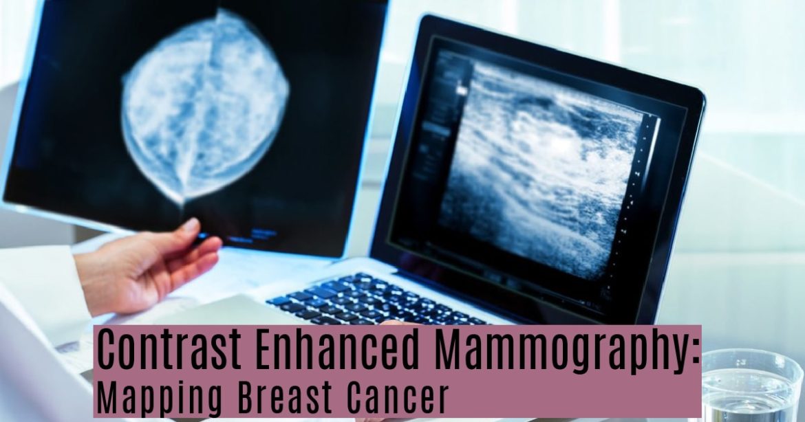
Contrast Enhanced Mammography Mapping Breast Cancer I Uva Radiology

Contrast Enhanced Mammography Mapping Breast Cancer I Uva Radiology Contrast enhanced spectral mammography (cem) is a specific type of mammogram exam that uses medical imaging contrast to create a special picture of the breast tissue. essentially, the image that cem creates is a composite of two images taken simultaneously—one showing the contrast, the other a regular mammogram. the contrast’s job is to. Contrast enhanced mammograms. make an appointment. for the charlottesville area: 434.243.4704. for culpeper: 540.321.3190. use the online form. if you have dense breast tissue or a high breast cancer risk, you need accurate, fast screening. with contrast enhanced mammograms (cem), we can discover cancers before standard mammograms.

Contrast Enhanced Mammography State Of The Art Radiology Contrast enhanced mammography (cem) has emerged as a viable alternative to contrast enhanced breast mri, and it may increase access to vascular imaging while reducing examination cost. intravenous iodinated contrast materials are used in cem to enhance the visualization of tumor neovascularity. after injection, imaging is performed with dual energy digital mammography, which helps provide a. Contrast enhanced mammography (cem) is an imaging technique that uses iodinated contrast medium to improve visualization of breast lesions and assessment of tumor neovascularity. through modifications in x ray energy, high and low energy images of the breast are combined to highlight areas of contrast medium pooling. the use of contrast material introduces different workflows, artifacts, and. Introduction. to date, full field digital mammography (ffdm) remains the primary imaging tool in breast cancer imaging worldwide. ffdm plays a pivotal role in breast cancer detection in clinical practice as well as in screening programmes. 1 however, ffdm is less accurate in females with dense breast tissue. 2,3 to resolve this issue, many technologies have been proposed as adjuncts to ffdm. To successfully implement contrast enhanced mammography into clinical practice, an understanding about the acquisition of images, image interpretation, and reporting of the spectrum of negative, benign, and malignant findings is essential. breast cancer remains the most common cancer diagnosis in women and was responsible for over 40,000 cancer.

Contrast Enhanced Mammography Techniques Current Results And Introduction. to date, full field digital mammography (ffdm) remains the primary imaging tool in breast cancer imaging worldwide. ffdm plays a pivotal role in breast cancer detection in clinical practice as well as in screening programmes. 1 however, ffdm is less accurate in females with dense breast tissue. 2,3 to resolve this issue, many technologies have been proposed as adjuncts to ffdm. To successfully implement contrast enhanced mammography into clinical practice, an understanding about the acquisition of images, image interpretation, and reporting of the spectrum of negative, benign, and malignant findings is essential. breast cancer remains the most common cancer diagnosis in women and was responsible for over 40,000 cancer. Purpose of review to provide an update on contrast enhanced mammography (cem) regarding current technique and interpretation, the performance of this modality versus conventional breast imaging modalities (mammography, ultrasound, and mri), existing clinical applications, potential challenges, and pitfalls. recent findings multiple studies have shown that the low energy, non contrast enhanced. Contrast enhanced mammography (cem) is a novel vascular based breast imaging technique which, like breast magnetic resonance imaging (mri), utilizes contrast media to depict neovascularity. angiogenesis, which drives breast cancer, recruits blood vessels that are more permeable to contrast, resulting in enhancement on imaging, sometimes before a discrete mass can be detected. a cem study uses.

Comments are closed.