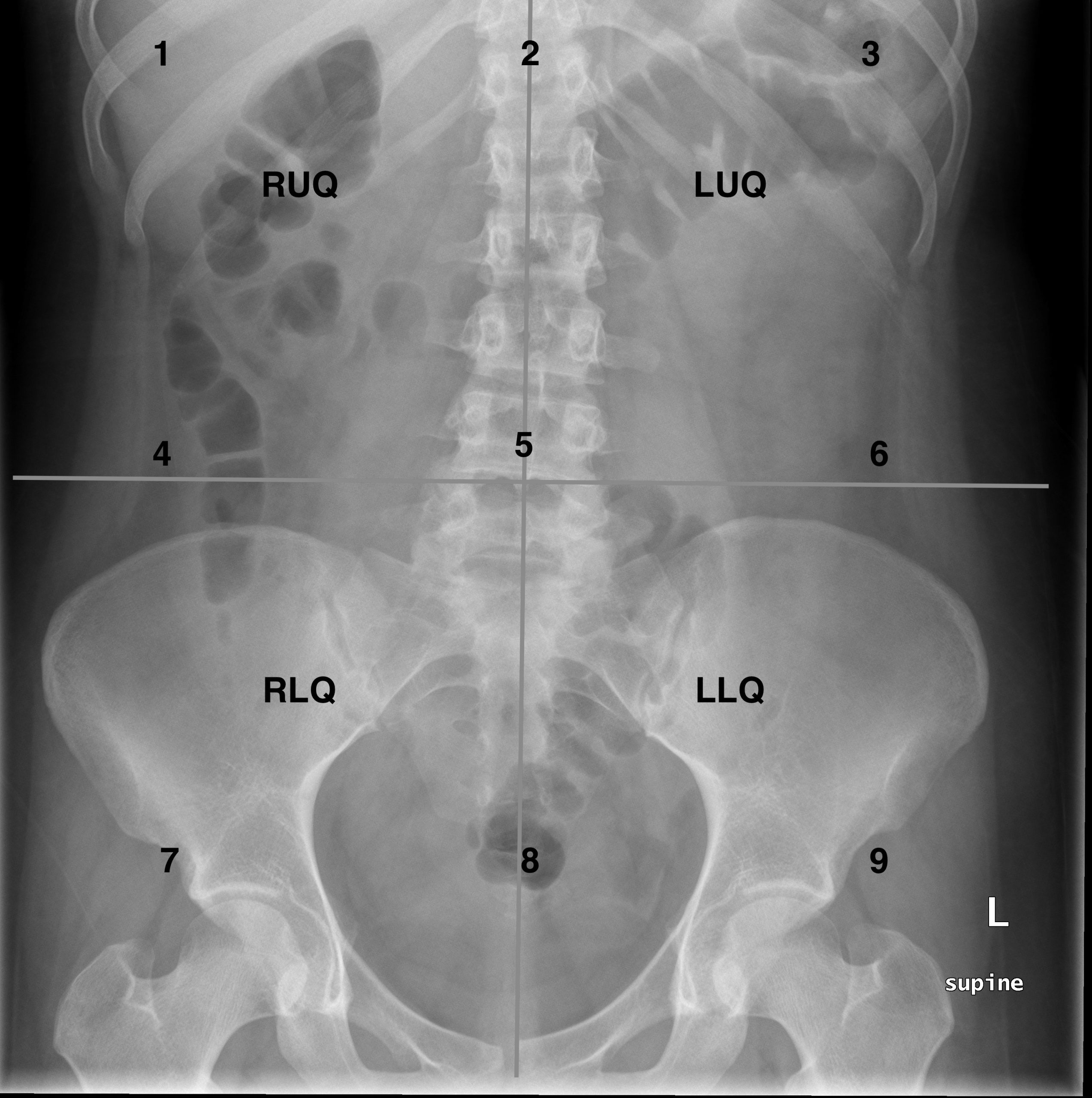
Ap Abdomen X Ray

Abdominal Radiographic Anatomy Wikiradiography Abdominal x ray (ap supine view) the ap supine abdominal radiograph can be performed as a standalone projection or as part of an acute abdominal series , depending on the clinical question posed, local protocol and the availability of other imaging modalities. Kub abdomen : patient supine and upright. the ap projection of the abdomen is sometimes called as kub xray, because in this projection the kidneys, ureters and bladder are included in the radiograph. it is also use as a preliminary evaluation radiograph or scot films for some of special procedure. when an ap projection of abdomen taken in.

Normal Abdominal Radiograph Radiology Case Radiopaedia Org The patient is supine, lying on his or her back, either on the x ray table (preferred) or a trolley. patients should be changed into a hospital gown, with radiopaque items removed (e.g. belts, zippers, buttons) the patient should be free from rotation; both shoulders and hips equidistant from the table trolley. Generally, plain radiograph examination of the abdomen comprises an ap supine and pa erect view, supplemented by a number of additional views as clinically indicated. nb: please note that in the uk, a single supine abdominal radiograph is the norm, i.e. erect abdominal x rays have not been routinely performed for decades 2. lateral decubitus view. Abdomen plane x ray (ap) supine and upright technique and positioning. Typical projections of an abdominal x ray include: anterior posterior (ap) supine; anterior posterior (ap) erect; exposure of image. assess the x ray to ensure the whole abdomen is visible from the level of the diaphragm to the pelvis. ensure the exposure is adequate to allow radiological assessment of both the small and large bowel. abdominal.

Ap Abdomen X Ray Abdomen plane x ray (ap) supine and upright technique and positioning. Typical projections of an abdominal x ray include: anterior posterior (ap) supine; anterior posterior (ap) erect; exposure of image. assess the x ray to ensure the whole abdomen is visible from the level of the diaphragm to the pelvis. ensure the exposure is adequate to allow radiological assessment of both the small and large bowel. abdominal. Abdominal x rays are usually acquired using an ap (anterior posterior) projection (x rays pass through the patient from front to back), with the patient positioned supine. erect chest x ray if perforation of the bowel is suspected then an erect chest x ray must be requested. Upright abdomen 1. 14 x 17 film 2. patient is in an ap erect position (allow time for free air to rise). 3. place top of cassette to the axilla (diaphragms must be demonstrated). 4. bucky 5. 40" sid 6. central ray: horizontal, parallel with the fluid level, even if patient isn't completely 90 degrees upright. 7. expiration. decubitus abdomen 1.

Approach To The Abdominal X Ray Axr вђ Undergraduate Diagnostic Abdominal x rays are usually acquired using an ap (anterior posterior) projection (x rays pass through the patient from front to back), with the patient positioned supine. erect chest x ray if perforation of the bowel is suspected then an erect chest x ray must be requested. Upright abdomen 1. 14 x 17 film 2. patient is in an ap erect position (allow time for free air to rise). 3. place top of cassette to the axilla (diaphragms must be demonstrated). 4. bucky 5. 40" sid 6. central ray: horizontal, parallel with the fluid level, even if patient isn't completely 90 degrees upright. 7. expiration. decubitus abdomen 1.

Abdomen Ap Supine View Radiology Reference Article Radiopaedia Org

Comments are closed.