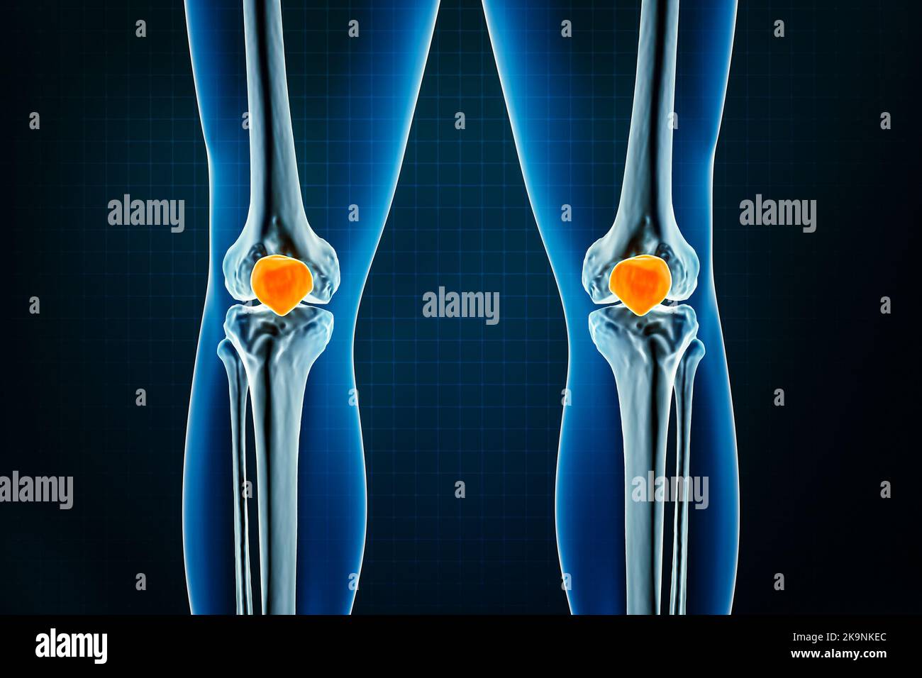
Anterior Or Front Close Up View Of The Patella Or Knee Cap Bone 3d

Anterior Or Front Close Up View Of The Patella Or Knee Cap Bone 3d The knee, also known as the tibiofemoral joint, is a synovial hinge joint formed between three bones: the femur, tibia, and patella. two rounded, convex processes (known as condyles) on the distal end of the femur meet two rounded, concave condyles at the proximal end of the tibia. the patella lies in front of the femur on the anterior surface. Knee. the knee is a complex joint that flexes, extends, and twists slightly from side to side. the knee is the meeting point of the femur (thigh bone) in the upper leg and the tibia (shinbone) in.

Patella Or Kneecap Bone X Ray Front Or Anterior View Osteology Of The Anatomy. the patella is the largest sesamoid bone in the body and it lies within the quadriceps tendon in front of the knee joint. the bone originates from multiple ossification centres that develop from the ages of three to six, which rapidly coalesce. the patella is a thick, flat, triangular bone with its apex pointing downwards. Knee joint (articulatio genu) the knee joint is a synovial joint that connects three bones; the femur, tibia and patella. it is a complex hinge joint composed of two articulations; the tibiofemoral joint and patellofemoral joint. the tibiofemoral joint is an articulation between the tibia and the femur, while the patellofemoral joint is an. 1. knee bone anatomy. the most basic component of knee joint anatomy are the bones which provide the structure to the knee. there are four knee bones that fit together to make two different knee joints: femur: the thigh bone. patella: the kneecap. tibia: the main shin bone. fibula: the outer shin bone. The patella is your kneecap. it’s the bone at the front of your knee joint. it’s the biggest bone in your body embedded in a tendon (a sesamoid bone). your patella helps your quadriceps muscle move your leg, protects your knee joint, and supports lots of important muscles, tendons and ligaments. traumas that hurt your knee are the most.

Patella Or Kneecap Bone X Ray Front Or Anterior View Osteology Of The 1. knee bone anatomy. the most basic component of knee joint anatomy are the bones which provide the structure to the knee. there are four knee bones that fit together to make two different knee joints: femur: the thigh bone. patella: the kneecap. tibia: the main shin bone. fibula: the outer shin bone. The patella is your kneecap. it’s the bone at the front of your knee joint. it’s the biggest bone in your body embedded in a tendon (a sesamoid bone). your patella helps your quadriceps muscle move your leg, protects your knee joint, and supports lots of important muscles, tendons and ligaments. traumas that hurt your knee are the most. Anatomy of the patellar region. patella is a thick, flat, triangular bone, having concave anterior and convex posterior surfaces. the posterior surface articulates with the femur and is marked by two shallow depressions or facets, medial and lateral. being triangular, it has a pointy end and three sides. the pointy end is the apex and the three. In this case, it’s the quadriceps femoris tendon, which places the patella just anterior to the femoral condyles. its superior edge is curved whereas the inferior aspect converges onto an apex giving it a kind of upside down teardrop shape. the posterior surface of the patella contains an oval articular surface close to its superior edge.

Comments are closed.