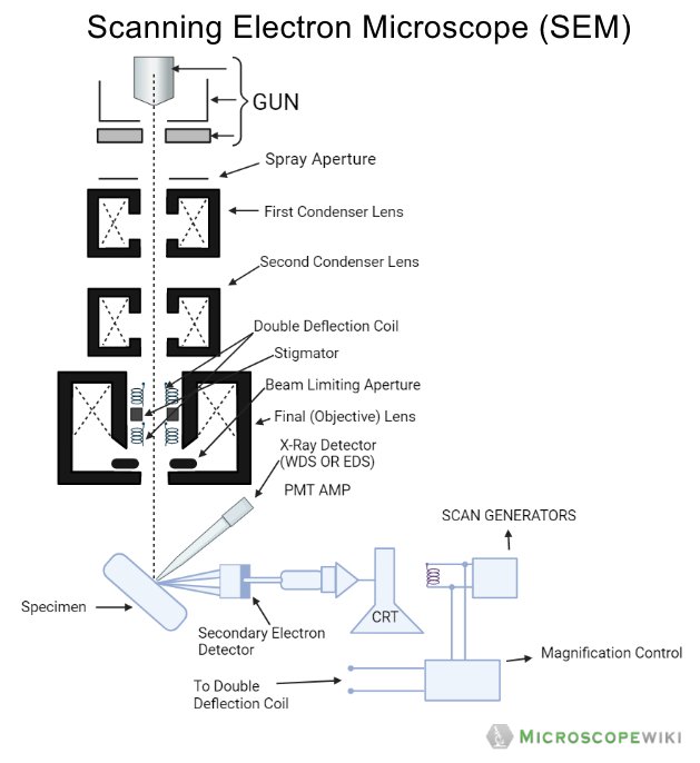
8 Schematic Drawing Of A The Typical Scanning Electron Microscope

8 Schematic Drawing Of A The Typical Scanning Electron Microscope A scanning electron microscope works on the principle of targeting a focused beam of electrons moving with high kinetic energy on a specimen. when these electrons scan the surface of the specimen, electrons are scattered and these secondary electrons that are slowed down on the impact of hitting the surface of the specimen are collected by a detector and these secondary electrons create the. Download scientific diagram | 8: schematic drawing of (a) the typical scanning electron microscope (sem) column [170], and (b) sample beam interactions within a sem. from publication.

8 Schematic Drawing Of A The Typical Scanning Electron Microscope The scanning electron microscope works on the principle of applying kinetic energy to produce signals on the interaction of the electrons. these electrons are secondary electrons, backscattered electrons, and diffracted backscattered electrons which are used to view crystallized elements and photons. secondary and backscattered electrons are. A brief description of each system follows: 1. vacuum system. a vacuum is required when using an electron beam because electrons will quickly disperse or scatter due to collisions with other molecules. 2. electron beam generation system. this system is found at the top of the microscope column (fig. 1). An account of the early history of scanning electron microscopy has been presented by mcmullan. [2] [3] although max knoll produced a photo with a 50 mm object field width showing channeling contrast by the use of an electron beam scanner, [4] it was manfred von ardenne who in 1937 invented [5] a microscope with high resolution by scanning a very small raster with a demagnified and finely. Stand the fundamentals of electron microscopy. the unaided eye can discriminate objects subtending about 1 60 ̊ visual angle, cor responding to a resolution. of ~0.1. mm (at the optimum viewing distance of25 cm). optical microscopy has the limit of resolution of ~2,000 Å by e.

Scanning Electron Microscope Sem Definition Images Uses An account of the early history of scanning electron microscopy has been presented by mcmullan. [2] [3] although max knoll produced a photo with a 50 mm object field width showing channeling contrast by the use of an electron beam scanner, [4] it was manfred von ardenne who in 1937 invented [5] a microscope with high resolution by scanning a very small raster with a demagnified and finely. Stand the fundamentals of electron microscopy. the unaided eye can discriminate objects subtending about 1 60 ̊ visual angle, cor responding to a resolution. of ~0.1. mm (at the optimum viewing distance of25 cm). optical microscopy has the limit of resolution of ~2,000 Å by e. Nthe goal of the sem is to scan a focused beam of primary electrons onto a sample, and to collect secondary electrons emitted from the sample to form an image. nmodern sems involve 5 main components. uan electron source (a.k.a electron gun) ufocusing and deflection optics (referred to as the column) ua specimen stage. Guide | scanning electron microscopy working principle 8 transmission electron microscopy (tem) in tem the accelerated electrons pass through the specimen. the transmitted ones then become focused as an enlarged image onto a fluorescent screen, which emits light when struck by these charged particles.

Schematic Of A Typical Scanning Electron Microscope A Vrogue Co Nthe goal of the sem is to scan a focused beam of primary electrons onto a sample, and to collect secondary electrons emitted from the sample to form an image. nmodern sems involve 5 main components. uan electron source (a.k.a electron gun) ufocusing and deflection optics (referred to as the column) ua specimen stage. Guide | scanning electron microscopy working principle 8 transmission electron microscopy (tem) in tem the accelerated electrons pass through the specimen. the transmitted ones then become focused as an enlarged image onto a fluorescent screen, which emits light when struck by these charged particles.

Comments are closed.