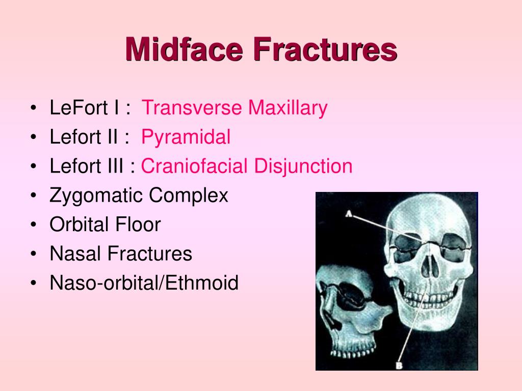
2 7 Midface Fractures

Midface Fractures Ento Key The aocmf craniomaxillofacial fracture classification system was developed in the form of a hierarchical three level system with increasing details and complexity. 1 within the midface, the level 2 system describes the location of the fractures within defined regions in the central and lateral midface with reference to classic le fort fractures and its analogs. 2. The commonly used classification is as follows: le fort type i. horizontal maxillary fracture, separating the teeth from the upper face. fracture line passes through the alveolar ridge, lateral nose and inferior wall of the maxillary sinus. also known as a guerin fracture. le fort type ii.

Ppt Midface Fractures Powerpoint Presentation Free Download Id 6370554 Abstract. facial fractures are a common source of emergency department consultations for the plastic surgeon. a working understanding of evaluation, assessment, management, and prevention of further injury when dealing with these fractures is vital. this two part series detailing the management of midface fractures serves as a guide for the. Concerning the anatomical site of fractures, it was explored that most common site of fractures is orbit (59, 25.72%) followed by fractures of maxilla (55, 24%) and zygomatic complex (35, 15.28%). Fractures of the midface, which includes the area from the superior orbital rim to the maxillary teeth, can cause irregularity in the smooth contour of the cheeks, malar eminences, zygomatic arch, or orbital rims. the le fort classification (see figure le fort classification of midface fractures) can be used to describe midface fractures. Level 2, a basic system for refined fracture location, is appropriate for all craniomaxillofacial (cmf) specialties referring to the anatomical regions and subregions of the midface, whereas the level 3 system is designed for assessing fracture morphology. the present article refers to the level 2 classification system for fractures of the.

Midfacial Fractures Oral Surgery B D S Fractures of the midface, which includes the area from the superior orbital rim to the maxillary teeth, can cause irregularity in the smooth contour of the cheeks, malar eminences, zygomatic arch, or orbital rims. the le fort classification (see figure le fort classification of midface fractures) can be used to describe midface fractures. Level 2, a basic system for refined fracture location, is appropriate for all craniomaxillofacial (cmf) specialties referring to the anatomical regions and subregions of the midface, whereas the level 3 system is designed for assessing fracture morphology. the present article refers to the level 2 classification system for fractures of the. Computed tomography is commonly used to evaluate patients with blunt facial trauma. with the high definition of the current scanners, even small fractures of the facial skeleton can be visualized. in complex midface injuries, it can be difficult for the radiologist to know which fractures are important to point out to the surgeon. an understanding of the anatomically relevant and surgically. The highest level 1 system distinguish four major anatomical units including the mandible (code 91), midface (code 92), skull base (code 93), and cranial vault (code 94). this tutorial presents the level 2 system for the midface unit that concentrates on the location of the fractures within defined regions in the central (upper, intermediate.

Comments are closed.